Movie
Movie Controller


+ Open data

- Basic information
| Entry |  | ||||||||||||||||||
|---|---|---|---|---|---|---|---|---|---|---|---|---|---|---|---|---|---|---|---|
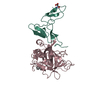
| Title | Cryo-EM structure of factor Va bound to activated protein C | ||||||||||||||||||
 Map data Map data | DeepEmhancer map. This map was used for model building | ||||||||||||||||||
 Sample Sample |
| ||||||||||||||||||
 Keywords Keywords | Coagulation / Activated Factor V / Activated Protein C / BLOOD CLOTTING | ||||||||||||||||||
| Function / homology |  Function and homology information Function and homology informationactivated protein C (thrombin-activated peptidase) / positive regulation of establishment of endothelial barrier / negative regulation of coagulation / response to vitamin K / platelet alpha granule / Cargo concentration in the ER / COPII-coated ER to Golgi transport vesicle / COPII-mediated vesicle transport / blood circulation / negative regulation of blood coagulation ...activated protein C (thrombin-activated peptidase) / positive regulation of establishment of endothelial barrier / negative regulation of coagulation / response to vitamin K / platelet alpha granule / Cargo concentration in the ER / COPII-coated ER to Golgi transport vesicle / COPII-mediated vesicle transport / blood circulation / negative regulation of blood coagulation / Transport of gamma-carboxylated protein precursors from the endoplasmic reticulum to the Golgi apparatus / : / Gamma-carboxylation of protein precursors / Removal of aminoterminal propeptides from gamma-carboxylated proteins / : / endoplasmic reticulum-Golgi intermediate compartment membrane / platelet alpha granule lumen / Cell surface interactions at the vascular wall / Post-translational protein phosphorylation / Golgi lumen / negative regulation of inflammatory response / Regulation of Insulin-like Growth Factor (IGF) transport and uptake by Insulin-like Growth Factor Binding Proteins (IGFBPs) / blood coagulation / Platelet degranulation / extracellular vesicle / endoplasmic reticulum lumen / copper ion binding / serine-type endopeptidase activity / calcium ion binding / negative regulation of apoptotic process / Golgi apparatus / endoplasmic reticulum / proteolysis / : / extracellular region / membrane / plasma membrane Similarity search - Function | ||||||||||||||||||
| Biological species |  Homo sapiens (human) Homo sapiens (human) | ||||||||||||||||||
| Method | single particle reconstruction / cryo EM / Resolution: 3.15 Å | ||||||||||||||||||
 Authors Authors | Mohammed BM / Basore K / Di Cera E | ||||||||||||||||||
| Funding support |  United States, 5 items United States, 5 items
| ||||||||||||||||||
 Citation Citation | Journal: Blood / Year: 2025 Title: Cryo-EM structure of coagulation factor Va bound to activated protein C. Authors: Bassem M Mohammed / Katherine Basore / Enrico Di Cera / Abstract: Coagulation factor Va (FVa) is the cofactor component of the prothrombinase complex required for rapid generation of thrombin from prothrombin in the penultimate step of the coagulation cascade. In ...Coagulation factor Va (FVa) is the cofactor component of the prothrombinase complex required for rapid generation of thrombin from prothrombin in the penultimate step of the coagulation cascade. In addition, FVa is a target for proteolytic inactivation by activated protein C (APC). Like other protein-protein interactions in the coagulation cascade, the FVa-APC interaction has long posed a challenge to structural biology and its molecular underpinnings remain unknown. A recent cryogenic electron microscopy (cryo-EM) structure of FVa has revealed the arrangement of its A1-A2-A3-C1-C2 domains and the environment of the sites of APC cleavage at R306 and R506. Here, we report the cryo-EM structure of the FVa-APC complex at 3.15 Å resolution in which the protease domain of APC engages R506 in the A2 domain of FVa through electrostatic interactions between positively charged residues in the 30-loop and 70-loop of APC and an electronegative surface of FVa. The auxiliary γ-carboxyglutamic acid and epidermal growth factor domains of APC are highly dynamic and point to solvent, without making contacts with FVa. Binding of APC displaces a large portion of the A2 domain of FVa and projects the 654VKCIPDDDEDSYEIFEP670 segment as a "latch," or exosite ligand, over the 70-loop of the enzyme. The latch induces a large conformational change of the autolysis loop of APC, which in turn promotes docking of R506 into the primary specificity pocket. The cryo-EM structure of the FVa-APC complex validates the bulk of existing biochemical data and offers molecular context for a key regulatory interaction of the coagulation cascade. | ||||||||||||||||||
| History |
|
- Structure visualization
| Supplemental images |
|---|

- Downloads & links
-EMDB archive
| Map data | emd_48481.map.gz | 247.7 MB | EMDB map data format | |
|---|---|---|---|---|
| Header (meta data) | emd-48481-v30.xmlemd-48481.xml | 38 KB 38 KB | Display Display | EMDB header |
| FSC (resolution estimation) | emd_48481_fsc.xml | 19.1 KB | Display | FSC data file |
| Images |  emd_48481.png emd_48481.png | 130.8 KB | ||
| Masks | emd_48481_msk_1.map | 282.6 MB | Mask map | |
| Filedesc metadata | emd-48481.cif.gz | 8.7 KB | ||
| Others | emd_48481_additional_1.map.gzemd_48481_additional_2.map.gzemd_48481_half_map_1.map.gzemd_48481_half_map_2.map.gz | 263.9 MB 264 MB 6.1 MB 6.1 MB | ||
| Archive directory |  http://ftp.pdbj.org/pub/emdb/structures/EMD-48481ftp://ftp.pdbj.org/pub/emdb/structures/EMD-48481 http://ftp.pdbj.org/pub/emdb/structures/EMD-48481ftp://ftp.pdbj.org/pub/emdb/structures/EMD-48481 | HTTPS FTP |
-Related structure data
| Related structure data |  9motMC 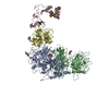 9movC C: citing same article ( M: atomic model generated by this map |
|---|---|
| Similar structure data |


-Links
| EMDB pages | EMDB (EBI/PDBe) / EMDataResource |
|---|---|
| Related items in Molecule of the Month |


-Map
| File | Download / File: emd_48481.map.gz / Format: CCP4 / Size: 282.6 MB / Type: IMAGE STORED AS FLOATING POINT NUMBER (4 BYTES) | ||||||||||||||||||||||||||||||||||||
|---|---|---|---|---|---|---|---|---|---|---|---|---|---|---|---|---|---|---|---|---|---|---|---|---|---|---|---|---|---|---|---|---|---|---|---|---|---|
| Annotation | DeepEmhancer map. This map was used for model building | ||||||||||||||||||||||||||||||||||||
| Projections & slices | Image control
Images are generated by Spider. | ||||||||||||||||||||||||||||||||||||
| Voxel size | X=Y=Z: 0.8701 Å | ||||||||||||||||||||||||||||||||||||
| Density |
| ||||||||||||||||||||||||||||||||||||
| Symmetry | Space group: 1 | ||||||||||||||||||||||||||||||||||||
| Details | EMDB XML:
|
 Z (Sec.)
 Y (Row.)
 X (Col.)


-Supplemental data
-Mask #1
| File | emd_48481_msk_1.map | ||||||||||||
|---|---|---|---|---|---|---|---|---|---|---|---|---|---|
| Projections & Slices |
| ||||||||||||
| Density Histograms |


-Additional map: 3D Flex map generated using custom mesh defining...
| File | emd_48481_additional_1.map | ||||||||||||
|---|---|---|---|---|---|---|---|---|---|---|---|---|---|
| Annotation | 3D Flex map generated using custom mesh defining each domain of the complex. | ||||||||||||
| Projections & Slices |
| ||||||||||||
| Density Histograms |


-Additional map: 3D Flex map generated using custom mesh defining...
| File | emd_48481_additional_2.map | ||||||||||||
|---|---|---|---|---|---|---|---|---|---|---|---|---|---|
| Annotation | 3D Flex map generated using custom mesh defining each domain of the complex. | ||||||||||||
| Projections & Slices |
| ||||||||||||
| Density Histograms |


-Half map: 3D flex half-maps
| File | emd_48481_half_map_1.map | ||||||||||||
|---|---|---|---|---|---|---|---|---|---|---|---|---|---|
| Annotation | 3D flex half-maps | ||||||||||||
| Projections & Slices |
| ||||||||||||
| Density Histograms |


-Half map: 3D flex half-maps
| File | emd_48481_half_map_2.map | ||||||||||||
|---|---|---|---|---|---|---|---|---|---|---|---|---|---|
| Annotation | 3D flex half-maps | ||||||||||||
| Projections & Slices |
| ||||||||||||
| Density Histograms |


- Sample components
-Entire : Complex of coagulation Factor Va and Activated Protein C
| Entire | Name: Complex of coagulation Factor Va and Activated Protein C |
|---|---|
| Components |
|
-Supramolecule #1: Complex of coagulation Factor Va and Activated Protein C
| Supramolecule | Name: Complex of coagulation Factor Va and Activated Protein C type: complex / ID: 1 / Parent: 0 / Macromolecule list: #1-#3 Details: Coagulation activated Factor V (FVa) [from plasma] complex with Activated Protein C (APC) [made recombinantly with Ser360Ala mutation]. |
|---|
-Supramolecule #2: Coagulation Factor Va
| Supramolecule | Name: Coagulation Factor Va / type: complex / ID: 2 / Parent: 1 / Macromolecule list: #1-#2 |
|---|---|
| Source (natural) | Organism: Homo sapiens (human) / Tissue: Blood |
-Supramolecule #3: Activated Protein C
| Supramolecule | Name: Activated Protein C / type: complex / ID: 3 / Parent: 1 / Macromolecule list: #3 |
|---|---|
| Source (natural) | Organism: Homo sapiens (human) |
-Macromolecule #1: Coagulation factor Va heavy chain
| Macromolecule | Name: Coagulation factor Va heavy chain / type: protein_or_peptide / ID: 1 / Number of copies: 1 / Enantiomer: LEVO |
|---|---|
| Source (natural) | Organism: Homo sapiens (human) / Tissue: blood |
| Molecular weight | Theoretical: 81.274391 KDa |
| Sequence | String: AQLRQFYVAA QGISWSYRPE PTNSSLNLSV TSFKKIVYRE YEPYFKKEKP QSTISGLLGP TLYAEVGDII KVHFKNKADK PLSIHPQGI RYSKLSEGAS YLDHTFPAEK MDDAVAPGRE YTYEWSISED SGPTHDDPPC LTHIYYSHEN LIEDFNSGLI G PLLICKKG ...String: AQLRQFYVAA QGISWSYRPE PTNSSLNLSV TSFKKIVYRE YEPYFKKEKP QSTISGLLGP TLYAEVGDII KVHFKNKADK PLSIHPQGI RYSKLSEGAS YLDHTFPAEK MDDAVAPGRE YTYEWSISED SGPTHDDPPC LTHIYYSHEN LIEDFNSGLI G PLLICKKG TLTEGGTQKT FDKQIVLLFA VFDESKSWSQ SSSLMYTVNG YVNGTMPDIT VCAHDHISWH LLGMSSGPEL FS IHFNGQV LEQNHHKVSA ITLVSATSTT ANMTVGPEGK WIISSLTPKH LQAGMQAYID IKNCPKKTRN LKKITREQRR HMK RWEYFI AAEEVIWDYA PVIPANMDKK YRSQHLDNFS NQIGKHYKKV MYTQYEDESF TKHTVNPNMK EDGILGPIIR AQVR DTLKI VFKNMASRPY SIYPHGVTFS PYEDEVNSSF TSGRNNTMIR AVQPGETYTY KWNILEFDEP TENDAQCLTR PYYSD VDIM RDIASGLIGL LLICKSRSLD RRGIQRAADI EQQAVFAVFD ENKSWYLEDN INKFCENPDE VKRDDPKFYE SNIMST ING YVPESITTLG FCFDDTVQWH FCSVGTQNEI LTIHFTGHSF IYGKRHEDTL TLFPMRGESV TVTMDNVGTW MLTSMNS SP RSKKLRLKFR DVKCIPDDDE DSYEIFEPPE STVMATRKMH DRLEPEDEES DADYDYQNRL AAALGIR UniProtKB: Coagulation factor V |
-Macromolecule #2: Coagulation factor Va light chain
| Macromolecule | Name: Coagulation factor Va light chain / type: protein_or_peptide / ID: 2 / Number of copies: 1 / Enantiomer: LEVO |
|---|---|
| Source (natural) | Organism: Homo sapiens (human) / Tissue: Blood |
| Molecular weight | Theoretical: 75.283008 KDa |
| Sequence | String: SNNGNRRNYY IAAEEISWDY SEFVQRETDI EDSDDIPEDT TYKKVVFRKY LDSTFTKRDP RGEYEEHLGI LGPIIRAEVD DVIQVRFKN LASRPYSLHA HGLSYEKSSE GKTYEDDSPE WFKEDNAVQP NSSYTYVWHA TERSGPESPG SACRAWAYYS A VNPEKDIH ...String: SNNGNRRNYY IAAEEISWDY SEFVQRETDI EDSDDIPEDT TYKKVVFRKY LDSTFTKRDP RGEYEEHLGI LGPIIRAEVD DVIQVRFKN LASRPYSLHA HGLSYEKSSE GKTYEDDSPE WFKEDNAVQP NSSYTYVWHA TERSGPESPG SACRAWAYYS A VNPEKDIH SGLIGPLLIC QKGILHKDSN MPMDMREFVL LFMTFDEKKS WYYEKKSRSS WRLTSSEMKK SHEFHAINGM IY SLPGLKM YEQEWVRLHL LNIGGSQDIH VVHFHGQTLL ENGNKQHQLG VWPLLPGSFK TLEMKASKPG WWLLNTEVGE NQR AGMQTP FLIMDRDCRM PMGLSTGIIS DSQIKASEFL GYWEPRLARL NNGGSYNAWS VEKLAAEFAS KPWIQVDMQK EVII TGIQT QGAKHYLKSC YTTEFYVAYS SNQINWQIFK GNSTRNVMYF NGNSDASTIK ENQFDPPIVA RYIRISPTRA YNRPT LRLE LQGCEVNGCS TPLGMENGKI ENKQITASSF KKSWWGDYWE PFRARLNAQG RVNAWQAKAN NNKQWLEIDL LKIKKI TAI ITQGCKSLSS EMYVKSYTIH YSEQGVEWKP YRLKSSMVDK IFEGNTNTKG HVKNFFNPPI ISRFIRVIPK TWNQSIA LR LELFGCDIY UniProtKB: Coagulation factor V |
-Macromolecule #3: Vitamin K-dependent protein C heavy chain
| Macromolecule | Name: Vitamin K-dependent protein C heavy chain / type: protein_or_peptide / ID: 3 / Number of copies: 1 / Enantiomer: LEVO |
|---|---|
| Source (natural) | Organism: Homo sapiens (human) / Tissue: Blood |
| Molecular weight | Theoretical: 26.949836 KDa |
| Recombinant expression | Organism:  Mesocricetus auratus (golden hamster) Mesocricetus auratus (golden hamster) |
| Sequence | String: LIDGKMTRRG DSPWQVVLLD SKKKLACGAV LIHPSWVLTA AHCMDESKKL LVRLGEYDLR RWEKWELDLD IKEVFVHPNY SKSTTDNDI ALLHLAQPAT LSQTIVPICL PDSGLAEREL NQAGQETLVT GWGYHSSREK EAKRNRTFVL NFIKIPVVPH N ECSEVMSN ...String: LIDGKMTRRG DSPWQVVLLD SKKKLACGAV LIHPSWVLTA AHCMDESKKL LVRLGEYDLR RWEKWELDLD IKEVFVHPNY SKSTTDNDI ALLHLAQPAT LSQTIVPICL PDSGLAEREL NQAGQETLVT GWGYHSSREK EAKRNRTFVL NFIKIPVVPH N ECSEVMSN MVSENMLCAG ILGDRQDACE GDAGGPMVAS FHGTWFLVGL VSWGEGCGLL HNYGVYTKVS RYLDWIHGHI RD UniProtKB: Vitamin K-dependent protein C |
-Macromolecule #4: 2-acetamido-2-deoxy-beta-D-glucopyranose
| Macromolecule | Name: 2-acetamido-2-deoxy-beta-D-glucopyranose / type: ligand / ID: 4 / Number of copies: 8 / Formula: NAG |
|---|---|
| Molecular weight | Theoretical: 221.208 Da |
| Chemical component information |  ChemComp-NAG: |
-Experimental details
-Structure determination
| Method | cryo EM |
|---|---|
 Processing Processing | single particle reconstruction |
| Aggregation state | particle |
-Sample preparation #1
| Preparation ID | 1 |
|---|---|
| Concentration | 0.1 mg/mL |
| Buffer | pH: 7.4 / Details: 20 mM HEPES, 150 mM NaCl, 5 mM CaCl2 |
| Grid | Model: Quantifoil R1.2/1.3 / Material: COPPER / Mesh: 300 / Support film - Film type ID: 1 / Support film - Material: CARBON / Support film - topology: HOLEY / Pretreatment - Type: GLOW DISCHARGE / Pretreatment - Time: 40 sec. / Pretreatment - Atmosphere: AIR / Details: Grids were coated with polylysine |
| Vitrification | Cryogen name: ETHANE / Chamber humidity: 100 % / Chamber temperature: 277.15 K / Instrument: FEI VITROBOT MARK IV |
| Details | 0.1 mg/mL |
-Sample preparation #2
| Preparation ID | 2 |
|---|---|
| Concentration | 0.1 mg/mL |
| Buffer | pH: 7.4 / Details: 20 mM HEPES, 150 mM NaCl, 5 mM CaCl2 |
| Grid | Model: Quantifoil R1.2/1.3 / Material: COPPER / Mesh: 300 / Support film - Film type ID: 1 / Support film - Material: CARBON / Support film - topology: HOLEY / Pretreatment - Type: GLOW DISCHARGE / Pretreatment - Time: 40 sec. / Pretreatment - Atmosphere: AIR / Details: Grids were coated with polylysine |
| Vitrification | Cryogen name: ETHANE / Chamber humidity: 100 % / Chamber temperature: 277.15 K / Instrument: FEI VITROBOT MARK IV |
-Sample preparation #3
| Preparation ID | 3 |
|---|---|
| Concentration | 0.1 mg/mL |
| Buffer | pH: 7.4 / Details: 20 mM HEPES, 150 mM NaCl, 5 mM CaCl2 |
| Grid | Model: Quantifoil R1.2/1.3 / Material: COPPER / Mesh: 300 / Support film - Film type ID: 1 / Support film - Material: CARBON / Support film - topology: HOLEY / Pretreatment - Type: GLOW DISCHARGE / Pretreatment - Time: 40 sec. / Pretreatment - Atmosphere: AIR / Details: Grids were coated with polylysine |
| Vitrification | Cryogen name: ETHANE / Chamber humidity: 100 % / Chamber temperature: 277.15 K / Instrument: FEI VITROBOT MARK IV |
- Electron microscopy
| Microscope | TFS KRIOS |
|---|---|
| Image recording | #0 - Image recording ID: 1 / #0 - Film or detector model: FEI FALCON IV (4k x 4k) / #0 - Detector mode: COUNTING / #0 - Digitization - Dimensions - Width: 4096 pixel / #0 - Digitization - Dimensions - Height: 4096 pixel / #0 - Number grids imaged: 1 / #0 - Number real images: 3193 / #0 - Average electron dose: 51.86 e/Å2 / #0 - Details: 30 degrees tilt / #1 - Image recording ID: 2 / #1 - Film or detector model: FEI FALCON IV (4k x 4k) / #1 - Digitization - Dimensions - Width: 4096 pixel / #1 - Digitization - Dimensions - Height: 4096 pixel / #1 - Number grids imaged: 1 / #1 - Number real images: 600 / #1 - Average electron dose: 46.6 e/Å2 / #2 - Image recording ID: 3 / #2 - Film or detector model: FEI FALCON IV (4k x 4k) / #2 - Digitization - Dimensions - Width: 4096 pixel / #2 - Digitization - Dimensions - Height: 4096 pixel / #2 - Number grids imaged: 1 / #2 - Number real images: 2655 / #2 - Average electron dose: 46.89 e/Å2 / #2 - Details: 30 degrees tilt / #3 - Image recording ID: 4 / #3 - Film or detector model: FEI FALCON IV (4k x 4k) / #3 - Digitization - Dimensions - Width: 4096 pixel / #3 - Digitization - Dimensions - Height: 4096 pixel / #3 - Number grids imaged: 1 / #3 - Number real images: 2379 / #3 - Average electron dose: 47.19 e/Å2 / #4 - Image recording ID: 5 / #4 - Film or detector model: FEI FALCON IV (4k x 4k) / #4 - Digitization - Dimensions - Width: 4096 pixel / #4 - Digitization - Dimensions - Height: 4096 pixel / #4 - Number grids imaged: 1 / #4 - Number real images: 420 / #4 - Average electron dose: 46.89 e/Å2 / #5 - Image recording ID: 6 / #5 - Film or detector model: FEI FALCON IV (4k x 4k) / #5 - Digitization - Dimensions - Width: 4096 pixel / #5 - Digitization - Dimensions - Height: 4096 pixel / #5 - Number grids imaged: 1 / #5 - Number real images: 3210 / #5 - Average electron dose: 52.8 e/Å2 |
| Electron beam | Acceleration voltage: 300 kV / Electron source:  FIELD EMISSION GUN FIELD EMISSION GUN |
| Electron optics | Illumination mode: FLOOD BEAM / Imaging mode: BRIGHT FIELD / Cs: 0.01 mm / Nominal defocus max: 2.5 µm / Nominal defocus min: 1.0 µm / Nominal magnification: 75000 |
| Sample stage | Specimen holder model: FEI TITAN KRIOS AUTOGRID HOLDER / Cooling holder cryogen: NITROGEN |
| Experimental equipment |  Model: Titan Krios / Image courtesy: FEI Company |
+Image processing

-Atomic model buiding 1
| Initial model |
| ||||||||
|---|---|---|---|---|---|---|---|---|---|
| Refinement | Space: REAL / Protocol: AB INITIO MODEL | ||||||||
| Output model | PDB-9mot: |