+ Open data
- Basic information
| Entry |  | ||||||||||||||||||
|---|---|---|---|---|---|---|---|---|---|---|---|---|---|---|---|---|---|---|---|
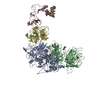
| Title | Cryo-EM structure of factor Va bound to activated protein C | ||||||||||||||||||
 Map data Map data | A1 domain local refined map | ||||||||||||||||||
 Sample Sample |
| ||||||||||||||||||
 Keywords Keywords | Coagulation / Activated Factor V / Activated Protein C / BLOOD CLOTTING | ||||||||||||||||||
| Biological species |  Homo sapiens (human) Homo sapiens (human) | ||||||||||||||||||
| Method | single particle reconstruction / cryo EM / Resolution: 3.0 Å | ||||||||||||||||||
 Authors Authors | Mohammed BM / Basore K / Di Cera E | ||||||||||||||||||
| Funding support |  United States, 5 items United States, 5 items
| ||||||||||||||||||
 Citation Citation |  Journal: Blood / Year: 2025 Journal: Blood / Year: 2025Title: Cryo-EM structure of coagulation factor Va bound to activated protein C. Authors: Bassem M Mohammed / Katherine Basore / Enrico Di Cera /  Abstract: Coagulation factor Va (FVa) is the cofactor component of the prothrombinase complex required for rapid generation of thrombin from prothrombin in the penultimate step of the coagulation cascade. In ...Coagulation factor Va (FVa) is the cofactor component of the prothrombinase complex required for rapid generation of thrombin from prothrombin in the penultimate step of the coagulation cascade. In addition, FVa is a target for proteolytic inactivation by activated protein C (APC). Like other protein-protein interactions in the coagulation cascade, the FVa-APC interaction has long posed a challenge to structural biology and its molecular underpinnings remain unknown. A recent cryogenic electron microscopy (cryo-EM) structure of FVa has revealed the arrangement of its A1-A2-A3-C1-C2 domains and the environment of the sites of APC cleavage at R306 and R506. Here, we report the cryo-EM structure of the FVa-APC complex at 3.15 Å resolution in which the protease domain of APC engages R506 in the A2 domain of FVa through electrostatic interactions between positively charged residues in the 30-loop and 70-loop of APC and an electronegative surface of FVa. The auxiliary γ-carboxyglutamic acid and epidermal growth factor domains of APC are highly dynamic and point to solvent, without making contacts with FVa. Binding of APC displaces a large portion of the A2 domain of FVa and projects the 654VKCIPDDDEDSYEIFEP670 segment as a "latch," or exosite ligand, over the 70-loop of the enzyme. The latch induces a large conformational change of the autolysis loop of APC, which in turn promotes docking of R506 into the primary specificity pocket. The cryo-EM structure of the FVa-APC complex validates the bulk of existing biochemical data and offers molecular context for a key regulatory interaction of the coagulation cascade. | ||||||||||||||||||
| History |
|
- Structure visualization
| Supplemental images |
|---|
- Downloads & links
-EMDB archive
| Map data |  emd_48465.map.gz emd_48465.map.gz | 141.4 MB |  EMDB map data format EMDB map data format | |
|---|---|---|---|---|
| Header (meta data) |  emd-48465-v30.xml emd-48465-v30.xml emd-48465.xml emd-48465.xml | 28 KB 28 KB | Display Display |  EMDB header EMDB header |
| FSC (resolution estimation) |  emd_48465_fsc.xml emd_48465_fsc.xml | 13.9 KB | Display |  FSC data file FSC data file |
| Images |  emd_48465.png emd_48465.png | 89.3 KB | ||
| Masks |  emd_48465_msk_1.map emd_48465_msk_1.map | 282.6 MB |  Mask map Mask map | |
| Filedesc metadata |  emd-48465.cif.gz emd-48465.cif.gz | 5.2 KB | ||
| Others |  emd_48465_additional_1.map.gz emd_48465_additional_1.map.gz emd_48465_additional_2.map.gz emd_48465_additional_2.map.gz emd_48465_half_map_1.map.gz emd_48465_half_map_1.map.gz emd_48465_half_map_2.map.gz emd_48465_half_map_2.map.gz | 447.9 KB 216.5 MB 262 MB 262 MB | ||
| Archive directory |  http://ftp.pdbj.org/pub/emdb/structures/EMD-48465 http://ftp.pdbj.org/pub/emdb/structures/EMD-48465 ftp://ftp.pdbj.org/pub/emdb/structures/EMD-48465 ftp://ftp.pdbj.org/pub/emdb/structures/EMD-48465 | HTTPS FTP |
-Related structure data
- Links
Links
| EMDB pages |  EMDB (EBI/PDBe) / EMDB (EBI/PDBe) /  EMDataResource EMDataResource |
|---|
- Map
Map
| File |  Download / File: emd_48465.map.gz / Format: CCP4 / Size: 282.6 MB / Type: IMAGE STORED AS FLOATING POINT NUMBER (4 BYTES) Download / File: emd_48465.map.gz / Format: CCP4 / Size: 282.6 MB / Type: IMAGE STORED AS FLOATING POINT NUMBER (4 BYTES) | ||||||||||||||||||||||||||||||||||||
|---|---|---|---|---|---|---|---|---|---|---|---|---|---|---|---|---|---|---|---|---|---|---|---|---|---|---|---|---|---|---|---|---|---|---|---|---|---|
| Annotation | A1 domain local refined map | ||||||||||||||||||||||||||||||||||||
| Projections & slices | Image control
Images are generated by Spider. | ||||||||||||||||||||||||||||||||||||
| Voxel size | X=Y=Z: 0.8701 Å | ||||||||||||||||||||||||||||||||||||
| Density |
| ||||||||||||||||||||||||||||||||||||
| Symmetry | Space group: 1 | ||||||||||||||||||||||||||||||||||||
| Details | EMDB XML:
|
-Supplemental data
-Mask #1
| File |  emd_48465_msk_1.map emd_48465_msk_1.map | ||||||||||||
|---|---|---|---|---|---|---|---|---|---|---|---|---|---|
| Projections & Slices |
| ||||||||||||
| Density Histograms |
-Additional map: Mask for DeepEmhancer
| File | emd_48465_additional_1.map | ||||||||||||
|---|---|---|---|---|---|---|---|---|---|---|---|---|---|
| Annotation | Mask for DeepEmhancer | ||||||||||||
| Projections & Slices |
| ||||||||||||
| Density Histograms |
-Additional map: DeepEmhancer sharpened map
| File | emd_48465_additional_2.map | ||||||||||||
|---|---|---|---|---|---|---|---|---|---|---|---|---|---|
| Annotation | DeepEmhancer sharpened map | ||||||||||||
| Projections & Slices |
| ||||||||||||
| Density Histograms |
-Half map: #1
| File | emd_48465_half_map_1.map | ||||||||||||
|---|---|---|---|---|---|---|---|---|---|---|---|---|---|
| Projections & Slices |
| ||||||||||||
| Density Histograms |
-Half map: #2
| File | emd_48465_half_map_2.map | ||||||||||||
|---|---|---|---|---|---|---|---|---|---|---|---|---|---|
| Projections & Slices |
| ||||||||||||
| Density Histograms |
- Sample components
-Entire : Complex of coagulation Factor Va and Activated Protein C
| Entire | Name: Complex of coagulation Factor Va and Activated Protein C |
|---|---|
| Components |
|
-Supramolecule #1: Complex of coagulation Factor Va and Activated Protein C
| Supramolecule | Name: Complex of coagulation Factor Va and Activated Protein C type: complex / ID: 1 / Parent: 0 Details: Coagulation activated Factor V (FVa) [from plasma] complex with Activated Protein C (APC) [made recombinantly with Ser360Ala mutation]. |
|---|
-Supramolecule #2: Coagulation Factor Va
| Supramolecule | Name: Coagulation Factor Va / type: complex / ID: 2 / Parent: 1 |
|---|---|
| Source (natural) | Organism:  Homo sapiens (human) / Tissue: Blood Homo sapiens (human) / Tissue: Blood |
-Supramolecule #3: Activated Protein C
| Supramolecule | Name: Activated Protein C / type: complex / ID: 3 / Parent: 1 |
|---|---|
| Source (natural) | Organism:  Homo sapiens (human) Homo sapiens (human) |
-Experimental details
-Structure determination
| Method | cryo EM |
|---|---|
 Processing Processing | single particle reconstruction |
| Aggregation state | particle |
- Sample preparation #1
| Preparation ID | 1 |
|---|---|
| Concentration | 0.1 mg/mL |
| Buffer | pH: 7.4 / Details: 20 mM HEPES, 150 mM NaCl, 5 mM CaCl2 |
| Grid | Model: Quantifoil R1.2/1.3 / Material: COPPER / Mesh: 300 / Support film - Film type ID: 1 / Support film - Material: CARBON / Support film - topology: HOLEY / Pretreatment - Type: GLOW DISCHARGE / Pretreatment - Time: 40 sec. / Pretreatment - Atmosphere: AIR / Details: Grids were coated with polylysine 0.1 mg/mL |
| Vitrification | Cryogen name: ETHANE / Chamber humidity: 100 % / Chamber temperature: 277.15 K / Instrument: FEI VITROBOT MARK IV |
| Details | 0.1 mg/mL |
- Sample preparation #2
| Preparation ID | 2 |
|---|---|
| Concentration | 0.1 mg/mL |
| Buffer | pH: 7.4 / Details: 20 mM HEPES, 150 mM NaCl, 5 mM CaCl2 |
| Grid | Model: Quantifoil R1.2/1.3 / Material: COPPER / Mesh: 300 / Support film - Film type ID: 1 / Support film - Material: CARBON / Support film - topology: HOLEY / Pretreatment - Type: GLOW DISCHARGE / Pretreatment - Time: 40 sec. / Pretreatment - Atmosphere: AIR / Details: Grids were coated with polylysine 0.1 mg/mL |
| Vitrification | Cryogen name: ETHANE / Chamber humidity: 100 % / Chamber temperature: 277.15 K / Instrument: FEI VITROBOT MARK IV |
- Sample preparation #3
| Preparation ID | 3 |
|---|---|
| Concentration | 0.1 mg/mL |
| Buffer | pH: 7.4 / Details: 20 mM HEPES, 150 mM NaCl, 5 mM CaCl2 |
| Grid | Model: Quantifoil R1.2/1.3 / Material: COPPER / Mesh: 300 / Support film - Film type ID: 1 / Support film - Material: CARBON / Support film - topology: HOLEY / Pretreatment - Type: GLOW DISCHARGE / Pretreatment - Time: 40 sec. / Pretreatment - Atmosphere: AIR / Details: Grids were coated with polylysine 0.1 mg/mL |
| Vitrification | Cryogen name: ETHANE / Chamber humidity: 100 % / Chamber temperature: 277.15 K / Instrument: FEI VITROBOT MARK IV |
- Electron microscopy
| Microscope | TFS KRIOS |
|---|---|
| Image recording | #0 - Image recording ID: 1 / #0 - Film or detector model: FEI FALCON IV (4k x 4k) / #0 - Detector mode: COUNTING / #0 - Digitization - Dimensions - Width: 4096 pixel / #0 - Digitization - Dimensions - Height: 4096 pixel / #0 - Number grids imaged: 1 / #0 - Number real images: 3193 / #0 - Average electron dose: 51.86 e/Å2 / #0 - Details: 30 degrees tilt / #1 - Image recording ID: 2 / #1 - Film or detector model: FEI FALCON IV (4k x 4k) / #1 - Digitization - Dimensions - Width: 4096 pixel / #1 - Digitization - Dimensions - Height: 4096 pixel / #1 - Number grids imaged: 1 / #1 - Number real images: 600 / #1 - Average electron dose: 46.6 e/Å2 / #2 - Image recording ID: 3 / #2 - Film or detector model: FEI FALCON IV (4k x 4k) / #2 - Digitization - Dimensions - Width: 4096 pixel / #2 - Digitization - Dimensions - Height: 4096 pixel / #2 - Number grids imaged: 1 / #2 - Number real images: 2655 / #2 - Average electron dose: 46.89 e/Å2 / #2 - Details: 30 degrees tilt / #3 - Image recording ID: 4 / #3 - Film or detector model: FEI FALCON IV (4k x 4k) / #3 - Digitization - Dimensions - Width: 4096 pixel / #3 - Digitization - Dimensions - Height: 4096 pixel / #3 - Number grids imaged: 1 / #3 - Number real images: 2379 / #3 - Average electron dose: 47.19 e/Å2 / #4 - Image recording ID: 5 / #4 - Film or detector model: FEI FALCON IV (4k x 4k) / #4 - Digitization - Dimensions - Width: 4096 pixel / #4 - Digitization - Dimensions - Height: 4096 pixel / #4 - Number grids imaged: 1 / #4 - Number real images: 420 / #4 - Average electron dose: 46.89 e/Å2 / #5 - Image recording ID: 6 / #5 - Film or detector model: FEI FALCON IV (4k x 4k) / #5 - Digitization - Dimensions - Width: 4096 pixel / #5 - Digitization - Dimensions - Height: 4096 pixel / #5 - Number grids imaged: 1 / #5 - Number real images: 3210 / #5 - Average electron dose: 52.8 e/Å2 |
| Electron beam | Acceleration voltage: 300 kV / Electron source:  FIELD EMISSION GUN FIELD EMISSION GUN |
| Electron optics | Illumination mode: FLOOD BEAM / Imaging mode: BRIGHT FIELD / Cs: 0.01 mm / Nominal defocus max: 2.5 µm / Nominal defocus min: 1.0 µm / Nominal magnification: 75000 |
| Sample stage | Specimen holder model: FEI TITAN KRIOS AUTOGRID HOLDER / Cooling holder cryogen: NITROGEN |
| Experimental equipment |  Model: Titan Krios / Image courtesy: FEI Company |
 Movie
Movie Controller


 Z (Sec.)
 Y (Row.)
 X (Col.)


