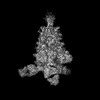
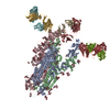
ジャーナル: Biochem Biophys Res Commun / 年: 2025 タイトル: Structure and dynamics of Alpha B.1.1.7 SARS-CoV-2 S-protein in complex with Fab of neutralizing antibody REGN10987. 著者: Milita V Kocharovskaya / Evgeny B Pichkur / Artem D Ivannikov / Daria D Kharlampieva / Ekaterina N Grafskaia / Ekaterina N Lyukmanova / Mikhail P Kirpichnikov / Zakhar O Shenkarev / 要旨: One of the approaches for treatment of COVID-19 is a use of neutralizing antibodies (nAbs). The study of the mechanisms by which nAbs recognize different strains of SARS-CoV-2 may facilitate the ...One of the approaches for treatment of COVID-19 is a use of neutralizing antibodies (nAbs). The study of the mechanisms by which nAbs recognize different strains of SARS-CoV-2 may facilitate the development of new drugs and vaccines against the coronavirus infection. In this work, we present the 3.1 Å resolution cryo-electron microscopy structure of a full-length trimeric spike-protein (S-protein) of the SARS-CoV-2 Alpha (B.1.1.7) variant in complex with the Fab of the REGN10987 nAb. In the complex, two receptor-binding domains (RBDs) of the S-protein were observed in the 'up' state, whereas third RBD was in the 'down' state. This distinguishes the obtained structure from the complex of Delta (B.1.617.2) S-protein with REGN10987-Fab, where only one RBD was in the 'up' state. Probably some of the substituted residues (K478T, A570D, and S982A) located at the interprotomer interfaces are responsible for the greater Alpha S-protein opening upon the REGN10987-Fab binding. The Fab identically binds to the RBD in the both 'up' and 'down' conformations. The RBD-Fab interaction interface was refined to a resolution of 3.6 Å. The antibody binds to the receptor-binding motif (RBM), which prevents the S-protein from the binding to its receptor, angiotensin-converting enzyme 2 (ACE-2). Comparison with the structures of the Wuhan (wild type) and Delta RBD variants in complex with REGN10987-Fab revealed that the N501Y and T478K/L452R mutations presented in the RBM of the Alpha and Delta variants, respectively, do not affect the mode of the RBD-Fab interaction.
 ムービー
ムービー コントローラー


 データを開く
 基本情報
基本情報 要素
要素 キーワード
キーワード 機能・相同性情報
 Severe acute respiratory syndrome coronavirus 2 (ウイルス)
 Homo sapiens (ヒト)
 データ登録者
 引用
引用
 構造の表示
構造の表示 ダウンロードとリンク
 その他のダウンロード
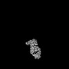
 PDBj
PDBj

 集合体
集合体
 試料調製
試料調製 電子顕微鏡撮影
 FIELD EMISSION GUN / 加速電圧: 300 kV / 照射モード: SPOT SCAN
 解析
解析