 Movie
Movie Controller

[English] 日本語
 Yorodumi
- PDB-9lyo: Alpha SARS-CoV-2 spike protein in complex with REGN10987 Fab homo... -
+ Open data

- Basic information
| Entry | Database: PDB / ID: 9lyo | ||||||
|---|---|---|---|---|---|---|---|
| Title | Alpha SARS-CoV-2 spike protein in complex with REGN10987 Fab homologue. | ||||||
 Components Components |
| ||||||
 Keywords Keywords | VIRAL PROTEIN | ||||||
| Function / homology |  Function and homology information Function and homology informationsymbiont-mediated disruption of host tissue / Maturation of spike protein / Translation of Structural Proteins / Virion Assembly and Release / host cell surface / host extracellular region / symbiont-mediated-mediated suppression of host tetherin activity / Induction of Cell-Cell Fusion / structural constituent of virion / positive regulation of viral entry into host cell ...symbiont-mediated disruption of host tissue / Maturation of spike protein / Translation of Structural Proteins / Virion Assembly and Release / host cell surface / host extracellular region / symbiont-mediated-mediated suppression of host tetherin activity / Induction of Cell-Cell Fusion / structural constituent of virion / positive regulation of viral entry into host cell / membrane fusion / host cell endoplasmic reticulum-Golgi intermediate compartment membrane / Attachment and Entry / entry receptor-mediated virion attachment to host cell / receptor-mediated virion attachment to host cell / host cell surface receptor binding / symbiont-mediated suppression of host innate immune response / endocytosis involved in viral entry into host cell / receptor ligand activity / fusion of virus membrane with host plasma membrane / fusion of virus membrane with host endosome membrane / viral envelope / symbiont entry into host cell / virion attachment to host cell / host cell plasma membrane / SARS-CoV-2 activates/modulates innate and adaptive immune responses / virion membrane / membrane / identical protein binding / plasma membrane Similarity search - Function | ||||||
| Biological species |  Homo sapiens (human) Homo sapiens (human)  Severe acute respiratory syndrome coronavirus 2 Severe acute respiratory syndrome coronavirus 2 | ||||||
| Method | ELECTRON MICROSCOPY / single particle reconstruction / cryo EM / Resolution: 3.07 Å | ||||||
 Authors Authors | Kocharovskaya, M.V. / Pichkur, E.B. / Shenkarev, Z.O. / Lyukmanova, E.N. | ||||||
| Funding support | 1items
| ||||||
 Citation Citation | Journal: Biochem Biophys Res Commun / Year: 2025 Title: Structure and dynamics of Alpha B.1.1.7 SARS-CoV-2 S-protein in complex with Fab of neutralizing antibody REGN10987. Authors: Milita V Kocharovskaya / Evgeny B Pichkur / Artem D Ivannikov / Daria D Kharlampieva / Ekaterina N Grafskaia / Ekaterina N Lyukmanova / Mikhail P Kirpichnikov / Zakhar O Shenkarev /  Abstract: One of the approaches for treatment of COVID-19 is a use of neutralizing antibodies (nAbs). The study of the mechanisms by which nAbs recognize different strains of SARS-CoV-2 may facilitate the ...One of the approaches for treatment of COVID-19 is a use of neutralizing antibodies (nAbs). The study of the mechanisms by which nAbs recognize different strains of SARS-CoV-2 may facilitate the development of new drugs and vaccines against the coronavirus infection. In this work, we present the 3.1 Å resolution cryo-electron microscopy structure of a full-length trimeric spike-protein (S-protein) of the SARS-CoV-2 Alpha (B.1.1.7) variant in complex with the Fab of the REGN10987 nAb. In the complex, two receptor-binding domains (RBDs) of the S-protein were observed in the 'up' state, whereas third RBD was in the 'down' state. This distinguishes the obtained structure from the complex of Delta (B.1.617.2) S-protein with REGN10987-Fab, where only one RBD was in the 'up' state. Probably some of the substituted residues (K478T, A570D, and S982A) located at the interprotomer interfaces are responsible for the greater Alpha S-protein opening upon the REGN10987-Fab binding. The Fab identically binds to the RBD in the both 'up' and 'down' conformations. The RBD-Fab interaction interface was refined to a resolution of 3.6 Å. The antibody binds to the receptor-binding motif (RBM), which prevents the S-protein from the binding to its receptor, angiotensin-converting enzyme 2 (ACE-2). Comparison with the structures of the Wuhan (wild type) and Delta RBD variants in complex with REGN10987-Fab revealed that the N501Y and T478K/L452R mutations presented in the RBM of the Alpha and Delta variants, respectively, do not affect the mode of the RBD-Fab interaction. | ||||||
| History |
|
- Structure visualization
| Structure viewer | Molecule: MolmilJmol/JSmol |
|---|
- Downloads & links
-Download
| PDBx/mmCIF format | 9lyo.cif.gz | 862 KB | Display | PDBx/mmCIF format |
|---|---|---|---|---|
| PDB format | pdb9lyo.ent.gz | 700.8 KB | Display | PDB format |
| PDBx/mmJSON format | 9lyo.json.gz | Tree view | PDBx/mmJSON format | |
| Others |  Other downloads Other downloads |
-Validation report
| Arichive directory | https://data.pdbj.org/pub/pdb/validation_reports/ly/9lyoftp://data.pdbj.org/pub/pdb/validation_reports/ly/9lyo | HTTPS FTP |
|---|
-Related structure data
| Related structure data | 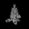 63513MC 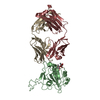 9lypC M: map data used to model this data C: citing same article ( |
|---|---|
| Similar structure data |
-Links
 PDBj
PDBj


- Assembly
| Deposited unit | 
|
|---|---|
| 1 |
|
-Components
| #1: Antibody | Mass: 23334.691 Da / Num. of mol.: 3 Source method: isolated from a genetically manipulated source Source: (gene. exp.) Homo sapiens (human) / Production host: Homo sapiens (human)#2: Antibody | Mass: 23792.617 Da / Num. of mol.: 3 Source method: isolated from a genetically manipulated source Source: (gene. exp.) Homo sapiens (human) / Production host: Homo sapiens (human)#3: Protein | Mass: 142662.797 Da / Num. of mol.: 3 Source method: isolated from a genetically manipulated source Source: (gene. exp.) Severe acute respiratory syndrome coronavirus 2Gene: S, 2 / Production host: Homo sapiens (human) / References: UniProt: P0DTC2#4: Polysaccharide | 2-acetamido-2-deoxy-beta-D-glucopyranose-(1-4)-2-acetamido-2-deoxy-beta-D-glucopyranose Source method: isolated from a genetically manipulated source #5: Sugar | ChemComp-NAG /   Type: D-saccharide, beta linking / Mass: 221.208 Da / Num. of mol.: 16 / Source method: obtained synthetically / Formula: C8H15NO6 Type: D-saccharide, beta linking / Mass: 221.208 Da / Num. of mol.: 16 / Source method: obtained synthetically / Formula: C8H15NO6Has ligand of interest | N | Has protein modification | Y | |
|---|
-Experimental details
-Experiment
| Experiment | Method: ELECTRON MICROSCOPY |
|---|---|
| EM experiment | Aggregation state: PARTICLE / 3D reconstruction method: single particle reconstruction |
- Sample preparation
| Component |
| ||||||||||||||||||||||||
|---|---|---|---|---|---|---|---|---|---|---|---|---|---|---|---|---|---|---|---|---|---|---|---|---|---|
| Source (natural) |
| ||||||||||||||||||||||||
| Source (recombinant) |
| ||||||||||||||||||||||||
| Buffer solution | pH: 7.5 | ||||||||||||||||||||||||
| Specimen | Embedding applied: NO / Shadowing applied: NO / Staining applied: NO / Vitrification applied: YES | ||||||||||||||||||||||||
| Vitrification | Cryogen name: ETHANE |
- Electron microscopy imaging
| Experimental equipment |  Model: Titan Krios / Image courtesy: FEI Company |
|---|---|
| Microscopy | Model: TFS KRIOS |
| Electron gun | Electron source:  FIELD EMISSION GUN / Accelerating voltage: 300 kV / Illumination mode: SPOT SCAN FIELD EMISSION GUN / Accelerating voltage: 300 kV / Illumination mode: SPOT SCAN |
| Electron lens | Mode: BRIGHT FIELD / Nominal defocus max: 1600 nm / Nominal defocus min: 800 nm |
| Image recording | Electron dose: 50 e/Å2 / Film or detector model: FEI FALCON IV (4k x 4k) |
- Processing
| EM software | Name: PHENIX / Version: 1.20.1_4487 / Category: model refinement |
|---|---|
| CTF correction | Type: PHASE FLIPPING AND AMPLITUDE CORRECTION |
| 3D reconstruction | Resolution: 3.07 Å / Resolution method: FSC 0.143 CUT-OFF / Num. of particles: 34988 / Symmetry type: POINT |
