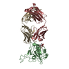
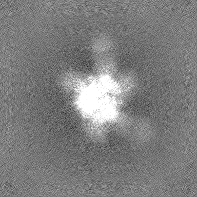
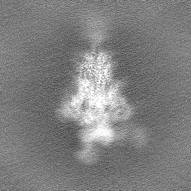
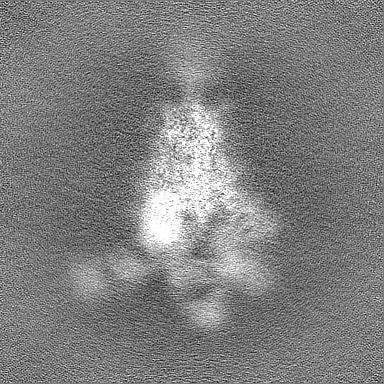
Journal: Biochem Biophys Res Commun / Year: 2025 Title: Structure and dynamics of Alpha B.1.1.7 SARS-CoV-2 S-protein in complex with Fab of neutralizing antibody REGN10987. Authors: Milita V Kocharovskaya / Evgeny B Pichkur / Artem D Ivannikov / Daria D Kharlampieva / Ekaterina N Grafskaia / Ekaterina N Lyukmanova / Mikhail P Kirpichnikov / Zakhar O Shenkarev / Abstract: One of the approaches for treatment of COVID-19 is a use of neutralizing antibodies (nAbs). The study of the mechanisms by which nAbs recognize different strains of SARS-CoV-2 may facilitate the ...One of the approaches for treatment of COVID-19 is a use of neutralizing antibodies (nAbs). The study of the mechanisms by which nAbs recognize different strains of SARS-CoV-2 may facilitate the development of new drugs and vaccines against the coronavirus infection. In this work, we present the 3.1 Å resolution cryo-electron microscopy structure of a full-length trimeric spike-protein (S-protein) of the SARS-CoV-2 Alpha (B.1.1.7) variant in complex with the Fab of the REGN10987 nAb. In the complex, two receptor-binding domains (RBDs) of the S-protein were observed in the 'up' state, whereas third RBD was in the 'down' state. This distinguishes the obtained structure from the complex of Delta (B.1.617.2) S-protein with REGN10987-Fab, where only one RBD was in the 'up' state. Probably some of the substituted residues (K478T, A570D, and S982A) located at the interprotomer interfaces are responsible for the greater Alpha S-protein opening upon the REGN10987-Fab binding. The Fab identically binds to the RBD in the both 'up' and 'down' conformations. The RBD-Fab interaction interface was refined to a resolution of 3.6 Å. The antibody binds to the receptor-binding motif (RBM), which prevents the S-protein from the binding to its receptor, angiotensin-converting enzyme 2 (ACE-2). Comparison with the structures of the Wuhan (wild type) and Delta RBD variants in complex with REGN10987-Fab revealed that the N501Y and T478K/L452R mutations presented in the RBM of the Alpha and Delta variants, respectively, do not affect the mode of the RBD-Fab interaction.
In the structure databanks used in Yorodumi, some data are registered as the other names, "COVID-19 virus" and "2019-nCoV". Here are the details of the virus and the list of structure data.
Jan 31, 2019. EMDB accession codes are about to change! (news from PDBe EMDB page)
EMDB accession codes are about to change! (news from PDBe EMDB page)
The allocation of 4 digits for EMDB accession codes will soon come to an end. Whilst these codes will remain in use, new EMDB accession codes will include an additional digit and will expand incrementally as the available range of codes is exhausted. The current 4-digit format prefixed with “EMD-” (i.e. EMD-XXXX) will advance to a 5-digit format (i.e. EMD-XXXXX), and so on. It is currently estimated that the 4-digit codes will be depleted around Spring 2019, at which point the 5-digit format will come into force.
The EM Navigator/Yorodumi systems omit the EMD- prefix.
Related info.:Q: What is EMD? / ID/Accession-code notation in Yorodumi/EM Navigator
Yorodumi is a browser for structure data from EMDB, PDB, SASBDB, etc.
This page is also the successor to EM Navigator detail page, and also detail information page/front-end page for Omokage search.
The word "yorodu" (or yorozu) is an old Japanese word meaning "ten thousand". "mi" (miru) is to see.
Related info.:EMDB / PDB / SASBDB / Comparison of 3 databanks / Yorodumi Search / Aug 31, 2016. New EM Navigator & Yorodumi / Yorodumi Papers / Jmol/JSmol / Function and homology information / Changes in new EM Navigator and Yorodumi
 Movie
Movie Controller

 Yorodumi
 Open data
 Basic information
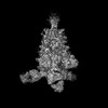
 Map data
 Sample
 Keywords
 Function and homology information
 Homo sapiens (human) /
 Severe acute respiratory syndrome coronavirus 2
 Authors
 Citation
 Structure visualization
 Downloads & links
 emd_63513.png
 http://ftp.pdbj.org/pub/emdb/structures/EMD-63513


 Z (Sec.)
 Y (Row.)
 X (Col.)


 Sample components
 Processing
 Electron microscopy
 FIELD EMISSION GUN


