[English] 日本語
 Yorodumi
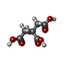
- PDB-9lsk: Cryo-EM structure of the Klebsiella pneumoniae CitS (citrate-boun... -
+ Open data
- Basic information
| Entry | Database: PDB / ID: 9lsk | |||||||||||||||||||||
|---|---|---|---|---|---|---|---|---|---|---|---|---|---|---|---|---|---|---|---|---|---|---|
| Title | Cryo-EM structure of the Klebsiella pneumoniae CitS (citrate-bound occluded state) | |||||||||||||||||||||
 Components Components | Citrate/sodium symporter | |||||||||||||||||||||
 Keywords Keywords | MEMBRANE PROTEIN / Disulfide-bridged diabody / cryo-electron microscopy (cryo-EM) / small protein imaging / structural marker / antibody engineering / protein nanotechnology | |||||||||||||||||||||
| Function / homology |  Function and homology information Function and homology informationcitrate metabolic process / : / symporter activity / sodium ion transport / metal ion binding / plasma membrane Similarity search - Function | |||||||||||||||||||||
| Biological species |  Klebsiella pneumoniae (bacteria) Klebsiella pneumoniae (bacteria) | |||||||||||||||||||||
| Method | ELECTRON MICROSCOPY / single particle reconstruction / cryo EM / Resolution: 2.9 Å | |||||||||||||||||||||
 Authors Authors | Kim, S. / Kim, J.W. / Park, J.G. / Lee, S.S. / Choi, S.H. / Lee, J.-O. / Jin, M.S. | |||||||||||||||||||||
| Funding support |  Korea, Republic Of, 3items Korea, Republic Of, 3items
| |||||||||||||||||||||
 Citation Citation |  Journal: Structure / Year: 2025 Journal: Structure / Year: 2025Title: Disulfide-stabilized diabodies enable near-atomic cryo-EM imaging of small proteins: A case study of the bacterial Na/citrate symporter CitS. Authors: Subin Kim / Ji Won Kim / Jun Gyou Park / Sang Soo Lee / Seung Hun Choi / Jie-Oh Lee / Mi Sun Jin /  Abstract: Diabodies are engineered antibody fragments with two antigen-binding Fv domains. Previously, we demonstrated that they are often highly flexible but can be rigidified by introducing a disulfide bond ...Diabodies are engineered antibody fragments with two antigen-binding Fv domains. Previously, we demonstrated that they are often highly flexible but can be rigidified by introducing a disulfide bond at the Fv interface. In this study, we explored the potential of disulfide-bridged, bispecific diabodies for near-atomic cryoelectron microscopy (cryo-EM) imaging of small proteins because they can predictably link target proteins to "structural marker" proteins. As a case study, we used the bacterial citrate transporter CitS as the target protein, and the horseshoe-shaped ectodomain of human Toll-like receptor 3 (TLR3) as the marker. We show that diabodies containing one or two disulfide bonds enabled the 3D reconstruction of CitS at resolutions of 3.3 Å and 3.1 Å, respectively. This resolution surpassed previous crystallographic results and allowed us to visualize the high-resolution structural features of the transporter. Our work expands the application of diabodies in structural biology to address a key limitation in the field. | |||||||||||||||||||||
| History |
|
- Structure visualization
| Structure viewer | Molecule:  Molmil Molmil Jmol/JSmol Jmol/JSmol |
|---|
- Downloads & links
- Download
| PDBx/mmCIF format |  9lsk.cif.gz 9lsk.cif.gz | 150.8 KB | Display |  PDBx/mmCIF format PDBx/mmCIF format |
|---|---|---|---|---|
| PDB format |  pdb9lsk.ent.gz pdb9lsk.ent.gz | 117.5 KB | Display |  PDB format PDB format |
| PDBx/mmJSON format |  9lsk.json.gz 9lsk.json.gz | Tree view |  PDBx/mmJSON format PDBx/mmJSON format | |
| Others |  Other downloads Other downloads |
-Validation report
| Summary document |  9lsk_validation.pdf.gz 9lsk_validation.pdf.gz | 1.4 MB | Display |  wwPDB validaton report wwPDB validaton report |
|---|---|---|---|---|
| Full document |  9lsk_full_validation.pdf.gz 9lsk_full_validation.pdf.gz | 1.4 MB | Display | |
| Data in XML |  9lsk_validation.xml.gz 9lsk_validation.xml.gz | 34 KB | Display | |
| Data in CIF |  9lsk_validation.cif.gz 9lsk_validation.cif.gz | 50.3 KB | Display | |
| Arichive directory |  https://data.pdbj.org/pub/pdb/validation_reports/ls/9lsk https://data.pdbj.org/pub/pdb/validation_reports/ls/9lsk ftp://data.pdbj.org/pub/pdb/validation_reports/ls/9lsk ftp://data.pdbj.org/pub/pdb/validation_reports/ls/9lsk | HTTPS FTP |
-Related structure data
| Related structure data |  63358MC  9lshC  9lsiC  9lsjC M: map data used to model this data C: citing same article ( |
|---|---|
| Similar structure data | Similarity search - Function & homology  F&H Search F&H Search |
- Links
Links
- Assembly
| Deposited unit | 
|
|---|---|
| 1 |
|
- Components
| #1: Protein | Mass: 47592.492 Da / Num. of mol.: 2 Source method: isolated from a genetically manipulated source Source: (gene. exp.)  Klebsiella pneumoniae (bacteria) / Gene: citS / Production host: Klebsiella pneumoniae (bacteria) / Gene: citS / Production host:  #2: Chemical | #3: Chemical | ChemComp-PLM / | Has ligand of interest | Y | Has protein modification | N | |
|---|
-Experimental details
-Experiment
| Experiment | Method: ELECTRON MICROSCOPY |
|---|---|
| EM experiment | Aggregation state: PARTICLE / 3D reconstruction method: single particle reconstruction |
- Sample preparation
| Component | Name: CitS / Type: COMPLEX / Entity ID: #1 / Source: RECOMBINANT |
|---|---|
| Source (natural) | Organism:  Klebsiella pneumoniae (bacteria) Klebsiella pneumoniae (bacteria) |
| Source (recombinant) | Organism:  |
| Buffer solution | pH: 7.5 |
| Specimen | Embedding applied: NO / Shadowing applied: NO / Staining applied: NO / Vitrification applied: YES |
| Vitrification | Cryogen name: ETHANE |
- Electron microscopy imaging
| Experimental equipment |  Model: Titan Krios / Image courtesy: FEI Company |
|---|---|
| Microscopy | Model: TFS KRIOS |
| Electron gun | Electron source:  FIELD EMISSION GUN / Accelerating voltage: 300 kV / Illumination mode: FLOOD BEAM FIELD EMISSION GUN / Accelerating voltage: 300 kV / Illumination mode: FLOOD BEAM |
| Electron lens | Mode: BRIGHT FIELD / Nominal defocus max: 1800 nm / Nominal defocus min: 800 nm |
| Image recording | Electron dose: 60 e/Å2 / Film or detector model: GATAN K3 BIOQUANTUM (6k x 4k) |
- Processing
| EM software | Name: PHENIX / Version: 1.18.2_3874 / Category: model refinement | ||||||||||||||||||||||||
|---|---|---|---|---|---|---|---|---|---|---|---|---|---|---|---|---|---|---|---|---|---|---|---|---|---|
| CTF correction | Type: PHASE FLIPPING AND AMPLITUDE CORRECTION | ||||||||||||||||||||||||
| 3D reconstruction | Resolution: 2.9 Å / Resolution method: FSC 0.143 CUT-OFF / Num. of particles: 235752 / Symmetry type: POINT | ||||||||||||||||||||||||
| Refinement | Stereochemistry target values: REAL-SPACE (WEIGHTED MAP SUM AT ATOM CENTERS) | ||||||||||||||||||||||||
| Refine LS restraints |
|
 Movie
Movie Controller


 PDBj
PDBj


