 Movie
Movie Controller

[English] 日本語
 Yorodumi
- EMDB-63358: Cryo-EM structure of the Klebsiella pneumoniae CitS (citrate-boun... -
+ Open data

- Basic information
| Entry |  | ||||||||||||
|---|---|---|---|---|---|---|---|---|---|---|---|---|---|
| Title | Cryo-EM structure of the Klebsiella pneumoniae CitS (citrate-bound occluded state) | ||||||||||||
 Map data Map data | |||||||||||||
 Sample Sample |
| ||||||||||||
 Keywords Keywords | Disulfide-bridged diabody / cryo-electron microscopy (cryo-EM) / small protein imaging / structural marker / antibody engineering / protein nanotechnology / MEMBRANE PROTEIN | ||||||||||||
| Function / homology |  Function and homology information Function and homology informationcitrate metabolic process / : / symporter activity / sodium ion transport / metal ion binding / plasma membrane Similarity search - Function | ||||||||||||
| Biological species |  Klebsiella pneumoniae (bacteria) Klebsiella pneumoniae (bacteria) | ||||||||||||
| Method | single particle reconstruction / cryo EM / Resolution: 2.9 Å | ||||||||||||
 Authors Authors | Kim S / Kim JW / Park JG / Lee SS / Choi SH / Lee J-O / Jin MS | ||||||||||||
| Funding support |  Korea, Republic Of, 3 items Korea, Republic Of, 3 items
| ||||||||||||
 Citation Citation | Journal: Structure / Year: 2025 Title: Disulfide-stabilized diabodies enable near-atomic cryo-EM imaging of small proteins: A case study of the bacterial Na/citrate symporter CitS. Authors: Subin Kim / Ji Won Kim / Jun Gyou Park / Sang Soo Lee / Seung Hun Choi / Jie-Oh Lee / Mi Sun Jin / Abstract: Diabodies are engineered antibody fragments with two antigen-binding Fv domains. Previously, we demonstrated that they are often highly flexible but can be rigidified by introducing a disulfide bond ...Diabodies are engineered antibody fragments with two antigen-binding Fv domains. Previously, we demonstrated that they are often highly flexible but can be rigidified by introducing a disulfide bond at the Fv interface. In this study, we explored the potential of disulfide-bridged, bispecific diabodies for near-atomic cryoelectron microscopy (cryo-EM) imaging of small proteins because they can predictably link target proteins to "structural marker" proteins. As a case study, we used the bacterial citrate transporter CitS as the target protein, and the horseshoe-shaped ectodomain of human Toll-like receptor 3 (TLR3) as the marker. We show that diabodies containing one or two disulfide bonds enabled the 3D reconstruction of CitS at resolutions of 3.3 Å and 3.1 Å, respectively. This resolution surpassed previous crystallographic results and allowed us to visualize the high-resolution structural features of the transporter. Our work expands the application of diabodies in structural biology to address a key limitation in the field. | ||||||||||||
| History |
|
- Structure visualization
| Supplemental images |
|---|

- Downloads & links
-EMDB archive
| Map data | emd_63358.map.gz | 30 MB | EMDB map data format | |
|---|---|---|---|---|
| Header (meta data) | emd-63358-v30.xmlemd-63358.xml | 18.2 KB 18.2 KB | Display Display | EMDB header |
| Images |  emd_63358.png emd_63358.png | 121.2 KB | ||
| Filedesc metadata | emd-63358.cif.gz | 6 KB | ||
| Others | emd_63358_half_map_1.map.gzemd_63358_half_map_2.map.gz | 55.3 MB 55.3 MB | ||
| Archive directory |  http://ftp.pdbj.org/pub/emdb/structures/EMD-63358ftp://ftp.pdbj.org/pub/emdb/structures/EMD-63358 http://ftp.pdbj.org/pub/emdb/structures/EMD-63358ftp://ftp.pdbj.org/pub/emdb/structures/EMD-63358 | HTTPS FTP |
-Related structure data
| Related structure data |  9lskMC  9lshC  9lsiC  9lsjC M: atomic model generated by this map C: citing same article ( |
|---|---|
| Similar structure data |


-Links
| EMDB pages | EMDB (EBI/PDBe) / EMDataResource |
|---|
-Map
| File | Download / File: emd_63358.map.gz / Format: CCP4 / Size: 59.6 MB / Type: IMAGE STORED AS FLOATING POINT NUMBER (4 BYTES) | ||||||||||||||||||||||||||||||||||||
|---|---|---|---|---|---|---|---|---|---|---|---|---|---|---|---|---|---|---|---|---|---|---|---|---|---|---|---|---|---|---|---|---|---|---|---|---|---|
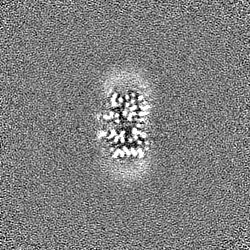
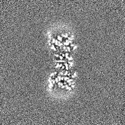
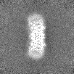
| Projections & slices | Image control
Images are generated by Spider. | ||||||||||||||||||||||||||||||||||||
| Voxel size | X=Y=Z: 0.8248 Å | ||||||||||||||||||||||||||||||||||||
| Density |
| ||||||||||||||||||||||||||||||||||||
| Symmetry | Space group: 1 | ||||||||||||||||||||||||||||||||||||
| Details | EMDB XML:
|
 Z (Sec.)
 Y (Row.)
 X (Col.)


-Supplemental data
-Half map: #1
| File | emd_63358_half_map_1.map | ||||||||||||
|---|---|---|---|---|---|---|---|---|---|---|---|---|---|
| Projections & Slices |
| ||||||||||||
| Density Histograms |


-Half map: #2
| File | emd_63358_half_map_2.map | ||||||||||||
|---|---|---|---|---|---|---|---|---|---|---|---|---|---|
| Projections & Slices |
| ||||||||||||
| Density Histograms |


- Sample components
-Entire : CitS
| Entire | Name: CitS |
|---|---|
| Components |
|
-Supramolecule #1: CitS
| Supramolecule | Name: CitS / type: complex / ID: 1 / Parent: 0 / Macromolecule list: #1 |
|---|---|
| Source (natural) | Organism: Klebsiella pneumoniae (bacteria) |
-Macromolecule #1: Citrate/sodium symporter
| Macromolecule | Name: Citrate/sodium symporter / type: protein_or_peptide / ID: 1 / Number of copies: 2 / Enantiomer: LEVO |
|---|---|
| Source (natural) | Organism: Klebsiella pneumoniae (bacteria) |
| Molecular weight | Theoretical: 47.592492 KDa |
| Recombinant expression | Organism: |
| Sequence | String: MTNMSQPPAT EKKGVSDLLG FKIFGMPLPL YAFALITLLL SHFYNALPTD IVGGFAIMFI IGAIFGEIGK RLPIFNKYIG GAPVMIFLV AAYFVYAGIF TQKEIDAISN VMDKSNFLNL FIAVLITGAI LSVNRRLLLK SLLGYIPTIL MGIVGASIFG I AIGLVFGI ...String: MTNMSQPPAT EKKGVSDLLG FKIFGMPLPL YAFALITLLL SHFYNALPTD IVGGFAIMFI IGAIFGEIGK RLPIFNKYIG GAPVMIFLV AAYFVYAGIF TQKEIDAISN VMDKSNFLNL FIAVLITGAI LSVNRRLLLK SLLGYIPTIL MGIVGASIFG I AIGLVFGI PVDRIMMLYV LPIMGGGNGA GAVPLSEIYH SVTGRSREEY YSTAIAILTI ANIFAIVFAA VLDIIGKKHT WL SGEGELV RKASFKVEED EKTGQITHRE TAVGLVLSTT CFLLAYVVAK KILPSIGGVA IHYFAWMVLI VAALNASGLC SPE IKAGAK RLSDFFSKQL LWVLMVGVGV CYTDLQEIIN AITFANVVIA AIIVIGAVLG AAIGGWLMGF FPIESAITAG LCMA NRGGS GDLEVLSACN RMNLISYAQI SSRLGGGIVL VIASIVFGMM I UniProtKB: Citrate/sodium symporter |
-Macromolecule #2: CITRIC ACID
| Macromolecule | Name: CITRIC ACID / type: ligand / ID: 2 / Number of copies: 2 / Formula: CIT |
|---|---|
| Molecular weight | Theoretical: 192.124 Da |
| Chemical component information |  ChemComp-CIT: |
-Macromolecule #3: PALMITIC ACID
| Macromolecule | Name: PALMITIC ACID / type: ligand / ID: 3 / Number of copies: 1 / Formula: PLM |
|---|---|
| Molecular weight | Theoretical: 256.424 Da |
| Chemical component information |  ChemComp-PLM: |
-Experimental details
-Structure determination
| Method | cryo EM |
|---|---|
 Processing Processing | single particle reconstruction |
| Aggregation state | particle |
-Sample preparation
| Buffer | pH: 7.5 |
|---|---|
| Vitrification | Cryogen name: ETHANE |
- Electron microscopy
| Microscope | TFS KRIOS |
|---|---|
| Image recording | Film or detector model: GATAN K3 BIOQUANTUM (6k x 4k) / Average electron dose: 60.0 e/Å2 |
| Electron beam | Acceleration voltage: 300 kV / Electron source:  FIELD EMISSION GUN FIELD EMISSION GUN |
| Electron optics | Illumination mode: FLOOD BEAM / Imaging mode: BRIGHT FIELD / Nominal defocus max: 1.8 µm / Nominal defocus min: 0.8 µm |
| Experimental equipment |  Model: Titan Krios / Image courtesy: FEI Company |
