 Movie
Movie Controller

[English] 日本語
 Yorodumi
- PDB-9l7e: Crystal structure of human kinesin-1 motor domain (G234A mutant) ... -
+ Open data

- Basic information
| Entry | Database: PDB / ID: 9l7e | ||||||
|---|---|---|---|---|---|---|---|
| Title | Crystal structure of human kinesin-1 motor domain (G234A mutant) in complex with ADP | ||||||
 Components Components | Kinesin-1 heavy chain | ||||||
 Keywords Keywords | MOTOR PROTEIN / Kinesin | ||||||
| Function / homology |  Function and homology information Function and homology informationregulation of modification of synapse structure, modulating synaptic transmission / cytolytic granule membrane / plus-end-directed vesicle transport along microtubule / cytoplasm organization / anterograde dendritic transport of neurotransmitter receptor complex / anterograde neuronal dense core vesicle transport / mitochondrion transport along microtubule / mitocytosis / retrograde neuronal dense core vesicle transport / anterograde axonal protein transport ...regulation of modification of synapse structure, modulating synaptic transmission / cytolytic granule membrane / plus-end-directed vesicle transport along microtubule / cytoplasm organization / anterograde dendritic transport of neurotransmitter receptor complex / anterograde neuronal dense core vesicle transport / mitochondrion transport along microtubule / mitocytosis / retrograde neuronal dense core vesicle transport / anterograde axonal protein transport / ciliary rootlet / lysosome localization / positive regulation of potassium ion transport / vesicle transport along microtubule / plus-end-directed microtubule motor activity / RHO GTPases activate KTN1 / Kinesins / kinesin complex / microtubule motor activity / centrosome localization / COPI-dependent Golgi-to-ER retrograde traffic / microtubule-based movement / stress granule disassembly / natural killer cell mediated cytotoxicity / synaptic vesicle transport / Insulin processing / postsynaptic cytosol / phagocytic vesicle / axon cytoplasm / dendrite cytoplasm / MHC class II antigen presentation / axon guidance / regulation of membrane potential / positive regulation of synaptic transmission, GABAergic / positive regulation of protein localization to plasma membrane / cellular response to type II interferon / centriolar satellite / Signaling by ALK fusions and activated point mutants / vesicle / nuclear membrane / microtubule binding / microtubule / cadherin binding / protein-containing complex binding / perinuclear region of cytoplasm / ATP hydrolysis activity / mitochondrion / ATP binding / identical protein binding / membrane / cytosol / cytoplasm Similarity search - Function | ||||||
| Biological species |  Homo sapiens (human) Homo sapiens (human) | ||||||
| Method |  X-RAY DIFFRACTION / SYNCHROTRON / MOLECULAR REPLACEMENT / Resolution: 2.4 Å X-RAY DIFFRACTION / SYNCHROTRON / MOLECULAR REPLACEMENT / Resolution: 2.4 Å | ||||||
 Authors Authors | Makino, T. / Miyazono, K. / Tanokura, M. / Tomishige, M. | ||||||
| Funding support |  France, 1items France, 1items
| ||||||
 Citation Citation | Journal: J Cell Biol / Year: 2025 Title: Tension-induced suppression of allosteric conformational changes coordinates kinesin-1 stepping. Authors: Tsukasa Makino / Ryo Kanada / Teppei Mori / Ken-Ichi Miyazono / Yuta Komori / Haruaki Yanagisawa / Shoji Takada / Masaru Tanokura / Masahide Kikkawa / Michio Tomishige /  Abstract: Kinesin-1 walks along microtubules by alternating ATP hydrolysis and movement of its two motor domains ("head"). The detached head preferentially binds to the forward tubulin-binding site after ATP ...Kinesin-1 walks along microtubules by alternating ATP hydrolysis and movement of its two motor domains ("head"). The detached head preferentially binds to the forward tubulin-binding site after ATP binds to the microtubule-bound head, but the mechanism preventing premature microtubule binding while the partner head awaits ATP remains unknown. Here, we examined the role of the neck linker, the segment connecting two heads, in this mechanism. Structural analyses of the nucleotide-free head revealed a bulge just ahead of the neck linker's base, creating an asymmetric constraint on its mobility. While the neck linker can stretch freely backward, it must navigate around this bulge to extend forward. We hypothesized that increased neck linker tension suppresses premature binding of the tethered head, which was supported by molecular dynamics simulations and single-molecule fluorescence assays. These findings demonstrate a tension-dependent allosteric mechanism that coordinates the movement of two heads, where neck linker tension modulates the allosteric conformational changes rather than directly affecting the nucleotide state. | ||||||
| History |
|
- Structure visualization
| Structure viewer | Molecule: MolmilJmol/JSmol |
|---|
- Downloads & links
-Download
| PDBx/mmCIF format | 9l7e.cif.gz | 137.8 KB | Display | PDBx/mmCIF format |
|---|---|---|---|---|
| PDB format | pdb9l7e.ent.gz | 105.2 KB | Display | PDB format |
| PDBx/mmJSON format | 9l7e.json.gz | Tree view | PDBx/mmJSON format | |
| Others |  Other downloads Other downloads |
-Validation report
| Summary document | 9l7e_validation.pdf.gz | 1.2 MB | Display | wwPDB validaton report |
|---|---|---|---|---|
| Full document | 9l7e_full_validation.pdf.gz | 1.2 MB | Display | |
| Data in XML | 9l7e_validation.xml.gz | 15.6 KB | Display | |
| Data in CIF | 9l7e_validation.cif.gz | 20.1 KB | Display | |
| Arichive directory | https://data.pdbj.org/pub/pdb/validation_reports/l7/9l7eftp://data.pdbj.org/pub/pdb/validation_reports/l7/9l7e | HTTPS FTP |
-Related structure data
| Related structure data | 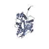 9l6kC  9l78C  9l7mC 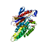 1bg2S C: citing same article ( S: Starting model for refinement |
|---|---|
| Similar structure data |

-Links
 PDBj
PDBj


- Assembly
| Deposited unit | 
| ||||||||
|---|---|---|---|---|---|---|---|---|---|
| 1 |
| ||||||||
| Unit cell |
|
-Components
| #1: Protein | Mass: 40016.918 Da / Num. of mol.: 1 / Mutation: G234A Source method: isolated from a genetically manipulated source Source: (gene. exp.) Homo sapiens (human) / Gene: KIF5B, KNS, KNS1 / Production host:  |
|---|---|
| #2: Chemical | ChemComp-MG /   Mass: 24.305 Da / Num. of mol.: 1 / Source method: obtained synthetically / Formula: Mg / Feature type: SUBJECT OF INVESTIGATION Mass: 24.305 Da / Num. of mol.: 1 / Source method: obtained synthetically / Formula: Mg / Feature type: SUBJECT OF INVESTIGATION |
| #3: Chemical | ChemComp-ADP /   Mass: 427.201 Da / Num. of mol.: 1 / Source method: obtained synthetically / Formula: C10H15N5O10P2 / Feature type: SUBJECT OF INVESTIGATION / Comment: ADP, energy-carrying molecule*YM Mass: 427.201 Da / Num. of mol.: 1 / Source method: obtained synthetically / Formula: C10H15N5O10P2 / Feature type: SUBJECT OF INVESTIGATION / Comment: ADP, energy-carrying molecule*YM |
| #4: Water | ChemComp-HOH /  Mass: 18.015 Da / Num. of mol.: 53 / Source method: isolated from a natural source / Formula: H2O Mass: 18.015 Da / Num. of mol.: 53 / Source method: isolated from a natural source / Formula: H2O |
| Has ligand of interest | Y |
| Has protein modification | N |
-Experimental details
-Experiment
| Experiment | Method: X-RAY DIFFRACTION / Number of used crystals: 1 |
|---|
- Sample preparation
| Crystal | Density Matthews: 2.31 Å3/Da / Density % sol: 46.76 % |
|---|---|
| Crystal grow | Temperature: 293 K / Method: vapor diffusion, sitting drop Details: 23% PEG 3350, 100mM Bis-Tris, pH 5.5, VAPOR DIFFUSION, SITTING DROP, temperature 293K |
-Data collection
| Diffraction | Mean temperature: 100 K / Serial crystal experiment: N |
|---|---|
| Diffraction source | Source: SYNCHROTRON / Site: Photon Factory / Beamline: AR-NW12A / Wavelength: 1 Å |
| Detector | Type: ADSC QUANTUM 210r / Detector: CCD / Date: Nov 9, 2006 |
| Radiation | Protocol: SINGLE WAVELENGTH / Monochromatic (M) / Laue (L): M / Scattering type: x-ray |
| Radiation wavelength | Wavelength: 1 Å / Relative weight: 1 |
| Reflection | Resolution: 2.4→20 Å / Num. obs: 14330 / % possible obs: 99.7 % / Redundancy: 4.2 % / Rmerge(I) obs: 0.052 / Net I/σ(I): 17.9 |
| Reflection shell | Resolution: 2.4→2.46 Å / Rmerge(I) obs: 0.412 / Mean I/σ(I) obs: 3.73 / Num. unique obs: 1056 |
- Processing
| Software |
| ||||||||||||||||||||||||||||||||||||||||||||||||||||||||||||||||||||||||||||||||||||||||||||||||||||||||||||||||||||||||||||||||||||||||||||||||||||||||||||||||||||||||||||||||||||||
|---|---|---|---|---|---|---|---|---|---|---|---|---|---|---|---|---|---|---|---|---|---|---|---|---|---|---|---|---|---|---|---|---|---|---|---|---|---|---|---|---|---|---|---|---|---|---|---|---|---|---|---|---|---|---|---|---|---|---|---|---|---|---|---|---|---|---|---|---|---|---|---|---|---|---|---|---|---|---|---|---|---|---|---|---|---|---|---|---|---|---|---|---|---|---|---|---|---|---|---|---|---|---|---|---|---|---|---|---|---|---|---|---|---|---|---|---|---|---|---|---|---|---|---|---|---|---|---|---|---|---|---|---|---|---|---|---|---|---|---|---|---|---|---|---|---|---|---|---|---|---|---|---|---|---|---|---|---|---|---|---|---|---|---|---|---|---|---|---|---|---|---|---|---|---|---|---|---|---|---|---|---|---|---|
| Refinement | Method to determine structure: MOLECULAR REPLACEMENT Starting model: PDB ENTRY 1BG2 Resolution: 2.4→19.55 Å / Cor.coef. Fo:Fc: 0.952 / Cor.coef. Fo:Fc free: 0.926 / SU B: 16.638 / SU ML: 0.189 / Cross valid method: THROUGHOUT / σ(F): 0 / ESU R Free: 0.261 / Stereochemistry target values: MAXIMUM LIKELIHOOD
| ||||||||||||||||||||||||||||||||||||||||||||||||||||||||||||||||||||||||||||||||||||||||||||||||||||||||||||||||||||||||||||||||||||||||||||||||||||||||||||||||||||||||||||||||||||||
| Solvent computation | Ion probe radii: 0.8 Å / Shrinkage radii: 0.8 Å / VDW probe radii: 1.4 Å / Solvent model: MASK | ||||||||||||||||||||||||||||||||||||||||||||||||||||||||||||||||||||||||||||||||||||||||||||||||||||||||||||||||||||||||||||||||||||||||||||||||||||||||||||||||||||||||||||||||||||||
| Displacement parameters | Biso mean: 45.25 Å2
| ||||||||||||||||||||||||||||||||||||||||||||||||||||||||||||||||||||||||||||||||||||||||||||||||||||||||||||||||||||||||||||||||||||||||||||||||||||||||||||||||||||||||||||||||||||||
| Refinement step | Cycle: LAST / Resolution: 2.4→19.55 Å
| ||||||||||||||||||||||||||||||||||||||||||||||||||||||||||||||||||||||||||||||||||||||||||||||||||||||||||||||||||||||||||||||||||||||||||||||||||||||||||||||||||||||||||||||||||||||
| Refine LS restraints |
|