 Movie
Movie Controller

[English] 日本語
 Yorodumi
- PDB-9j5c: ESTS1 phthalate ester degrading esterase from Sulfobacillus acido... -
+ Open data

- Basic information
| Entry | Database: PDB / ID: 9j5c | ||||||
|---|---|---|---|---|---|---|---|
| Title | ESTS1 phthalate ester degrading esterase from Sulfobacillus acidophilus in complex with diethylhexyl phthalate | ||||||
 Components Components | Alpha/beta hydrolase fold-3 domain-containing protein | ||||||
 Keywords Keywords | HYDROLASE / ESTS1 / phthalate ester degrading esterase / diethylhexyl phthalate | ||||||
| Function / homology |  Function and homology information Function and homology information | ||||||
| Biological species |  Sulfobacillus acidophilus DSM 10332 (bacteria) Sulfobacillus acidophilus DSM 10332 (bacteria) | ||||||
| Method |  X-RAY DIFFRACTION / MOLECULAR REPLACEMENT / Resolution: 2.7 Å X-RAY DIFFRACTION / MOLECULAR REPLACEMENT / Resolution: 2.7 Å | ||||||
 Authors Authors | Verma, S. / Kumar, P. | ||||||
| Funding support |  India, 1items India, 1items
| ||||||
 Citation Citation | Journal: Structure / Year: 2025 Title: Mechanistic and structural insights into EstS1 esterase: A potent broad-spectrum phthalate diester degrading enzyme. Authors: Verma, S. / Choudhary, S. / Amith Kumar, K. / Mahto, J.K. / Vamsi K, A.K. / Mishra, I. / Prakash, V.B. / Sircar, D. / Tomar, S. / Kumar Sharma, A. / Singla, J. / Kumar, P. | ||||||
| History |
|
- Structure visualization
| Structure viewer | Molecule: MolmilJmol/JSmol |
|---|
- Downloads & links
-Download
| PDBx/mmCIF format | 9j5c.cif.gz | 161 KB | Display | PDBx/mmCIF format |
|---|---|---|---|---|
| PDB format | pdb9j5c.ent.gz | 108.9 KB | Display | PDB format |
| PDBx/mmJSON format | 9j5c.json.gz | Tree view | PDBx/mmJSON format | |
| Others |  Other downloads Other downloads |
-Validation report
| Arichive directory | https://data.pdbj.org/pub/pdb/validation_reports/j5/9j5cftp://data.pdbj.org/pub/pdb/validation_reports/j5/9j5c | HTTPS FTP |
|---|
-Related structure data
| Related structure data |  8w98C  8ze9C  9j1eC 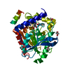 9j1gC  9j1vC  9j57C  9j58C  9j59C  9j5aC  9j5bC  9j5dC C: citing same article ( |
|---|---|
| Similar structure data |
-Links
 PDBj
PDBj- Assembly
| Deposited unit | 
| ||||||||
|---|---|---|---|---|---|---|---|---|---|
| 1 |
| ||||||||
| Unit cell |
|
-Components
| #1: Protein | Mass: 34176.836 Da / Num. of mol.: 1 Source method: isolated from a genetically manipulated source Source: (gene. exp.) Sulfobacillus acidophilus DSM 10332 (bacteria)Gene: Sulac_0033 / Production host: | ||||||
|---|---|---|---|---|---|---|---|
| #2: Chemical | Mass: 390.556 Da / Num. of mol.: 2 / Source method: obtained synthetically / Formula: C24H38O4 / Feature type: SUBJECT OF INVESTIGATION #3: Water | ChemComp-HOH / |  Mass: 18.015 Da / Num. of mol.: 84 / Source method: isolated from a natural source / Formula: H2O Mass: 18.015 Da / Num. of mol.: 84 / Source method: isolated from a natural source / Formula: H2OHas ligand of interest | Y | Has protein modification | N | |
-Experimental details
-Experiment
| Experiment | Method: X-RAY DIFFRACTION / Number of used crystals: 1 |
|---|
- Sample preparation
| Crystal | Density Matthews: 2.278 Å3/Da / Density % sol: 46.049 % |
|---|---|
| Crystal grow | Temperature: 293.15 K / Method: vapor diffusion / pH: 7 Details: Sodium malonate, 0.1 M HEPES (pH 7.0), Jeffamine ED-2001 |
-Data collection
| Diffraction | Mean temperature: 100 K / Serial crystal experiment: N |
|---|---|
| Diffraction source | Source: ROTATING ANODE / Type: RIGAKU MICROMAX-007 HF / Wavelength: 1.54 Å |
| Detector | Type: RIGAKU HyPix-6000HE / Detector: PIXEL / Date: Jan 14, 2023 |
| Radiation | Protocol: SINGLE WAVELENGTH / Monochromatic (M) / Laue (L): M / Scattering type: x-ray |
| Radiation wavelength | Wavelength: 1.54 Å / Relative weight: 1 |
| Reflection | Resolution: 2.7→25.64 Å / Num. obs: 8440 / % possible obs: 99.9 % / Redundancy: 3.06 % / Rrim(I) all: 0.43 / Net I/σ(I): 15.5 |
| Reflection shell | Resolution: 2.7→2.83 Å / Mean I/σ(I) obs: 3.7 / Num. unique obs: 8440 / Rrim(I) all: 1 |
- Processing
| Software |
| ||||||||||||||||||||||||||||||||||||||||||||||||||||||||||||||||||||||||||||||||||||||||||||||||||||||||||||||||||||||||||||||||||||||||||||||||||||||||||||||||
|---|---|---|---|---|---|---|---|---|---|---|---|---|---|---|---|---|---|---|---|---|---|---|---|---|---|---|---|---|---|---|---|---|---|---|---|---|---|---|---|---|---|---|---|---|---|---|---|---|---|---|---|---|---|---|---|---|---|---|---|---|---|---|---|---|---|---|---|---|---|---|---|---|---|---|---|---|---|---|---|---|---|---|---|---|---|---|---|---|---|---|---|---|---|---|---|---|---|---|---|---|---|---|---|---|---|---|---|---|---|---|---|---|---|---|---|---|---|---|---|---|---|---|---|---|---|---|---|---|---|---|---|---|---|---|---|---|---|---|---|---|---|---|---|---|---|---|---|---|---|---|---|---|---|---|---|---|---|---|---|---|---|
| Refinement | Method to determine structure: MOLECULAR REPLACEMENT / Resolution: 2.7→23.45 Å / Cor.coef. Fo:Fc: 0.887 / Cor.coef. Fo:Fc free: 0.75 / SU B: 41.144 / SU ML: 0.384 / Cross valid method: FREE R-VALUE / ESU R Free: 0.448 Details: Hydrogens have been added in their riding positions
| ||||||||||||||||||||||||||||||||||||||||||||||||||||||||||||||||||||||||||||||||||||||||||||||||||||||||||||||||||||||||||||||||||||||||||||||||||||||||||||||||
| Solvent computation | Ion probe radii: 0.8 Å / Shrinkage radii: 0.8 Å / VDW probe radii: 1.2 Å / Solvent model: MASK BULK SOLVENT | ||||||||||||||||||||||||||||||||||||||||||||||||||||||||||||||||||||||||||||||||||||||||||||||||||||||||||||||||||||||||||||||||||||||||||||||||||||||||||||||||
| Displacement parameters | Biso mean: 5 Å2
| ||||||||||||||||||||||||||||||||||||||||||||||||||||||||||||||||||||||||||||||||||||||||||||||||||||||||||||||||||||||||||||||||||||||||||||||||||||||||||||||||
| Refinement step | Cycle: LAST / Resolution: 2.7→23.45 Å
| ||||||||||||||||||||||||||||||||||||||||||||||||||||||||||||||||||||||||||||||||||||||||||||||||||||||||||||||||||||||||||||||||||||||||||||||||||||||||||||||||
| Refine LS restraints |
| ||||||||||||||||||||||||||||||||||||||||||||||||||||||||||||||||||||||||||||||||||||||||||||||||||||||||||||||||||||||||||||||||||||||||||||||||||||||||||||||||
| LS refinement shell |
| ||||||||||||||||||||||||||||||||||||||||||||||||||||||||||||||||||||||||||||||||||||||||||||||||||||||||||||||||||||||||||||||||||||||||||||||||||||||||||||||||
| Refinement TLS params. | Method: refined / Origin x: -26.3822 Å / Origin y: -30.2149 Å / Origin z: -0.3103 Å
| ||||||||||||||||||||||||||||||||||||||||||||||||||||||||||||||||||||||||||||||||||||||||||||||||||||||||||||||||||||||||||||||||||||||||||||||||||||||||||||||||
| Refinement TLS group |
|
