 Movie
Movie Controller

[English] 日本語
 Yorodumi
- PDB-8ze9: ESTS1 phthalate ester degrading esterase from Sulfobacillus acido... -
+ Open data

- Basic information
| Entry | Database: PDB / ID: 8ze9 | ||||||
|---|---|---|---|---|---|---|---|
| Title | ESTS1 phthalate ester degrading esterase from Sulfobacillus acidophilus S154A mutant in complex with diethylhexyl phthalate at 2.4A | ||||||
 Components Components | Triacylglycerol lipase | ||||||
 Keywords Keywords | HYDROLASE / Esterase / Phthalate Ester degrading / ESTS1 / HYDROLASE Diethylhexyl phthalate | ||||||
| Function / homology |  Function and homology information Function and homology information | ||||||
| Biological species |  Sulfobacillus acidophilus DSM 10332 (bacteria) Sulfobacillus acidophilus DSM 10332 (bacteria) | ||||||
| Method |  X-RAY DIFFRACTION / MOLECULAR REPLACEMENT / Resolution: 2.4 Å X-RAY DIFFRACTION / MOLECULAR REPLACEMENT / Resolution: 2.4 Å | ||||||
 Authors Authors | Verma, S. / Choudhary, S. / Kumar, P. | ||||||
| Funding support |  India, 1items India, 1items
| ||||||
 Citation Citation | Journal: Structure / Year: 2025 Title: Mechanistic and structural insights into EstS1 esterase: A potent broad-spectrum phthalate diester degrading enzyme. Authors: Verma, S. / Choudhary, S. / Amith Kumar, K. / Mahto, J.K. / Vamsi K, A.K. / Mishra, I. / Prakash, V.B. / Sircar, D. / Tomar, S. / Kumar Sharma, A. / Singla, J. / Kumar, P. | ||||||
| History |
|
- Structure visualization
| Structure viewer | Molecule: MolmilJmol/JSmol |
|---|
- Downloads & links
-Download
| PDBx/mmCIF format | 8ze9.cif.gz | 77.8 KB | Display | PDBx/mmCIF format |
|---|---|---|---|---|
| PDB format | pdb8ze9.ent.gz | 54.4 KB | Display | PDB format |
| PDBx/mmJSON format | 8ze9.json.gz | Tree view | PDBx/mmJSON format | |
| Others |  Other downloads Other downloads |
-Validation report
| Arichive directory | https://data.pdbj.org/pub/pdb/validation_reports/ze/8ze9ftp://data.pdbj.org/pub/pdb/validation_reports/ze/8ze9 | HTTPS FTP |
|---|
-Related structure data
| Related structure data |  8w98C  9j1eC 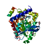 9j1gC  9j1vC  9j57C  9j58C  9j59C  9j5aC  9j5bC  9j5cC  9j5dC  8ioq 8ip7 8iph 8isd 8isl 8ix4 8izo 8izx 8j3d 8j3e C: citing same article ( |
|---|---|
| Similar structure data |
-Links
 PDBj
PDBj- Assembly
| Deposited unit | 
| ||||||||
|---|---|---|---|---|---|---|---|---|---|
| 1 |
| ||||||||
| Unit cell |
|
-Components
| #1: Protein | Mass: 33331.953 Da / Num. of mol.: 1 / Mutation: S154A Source method: isolated from a genetically manipulated source Source: (gene. exp.) Sulfobacillus acidophilus DSM 10332 (bacteria)Gene: Sulac_0033 / Production host: | ||||||
|---|---|---|---|---|---|---|---|
| #2: Chemical | ChemComp-TKU / ~{ Mass: 390.556 Da / Num. of mol.: 1 / Source method: obtained synthetically / Formula: C24H38O4 / Feature type: SUBJECT OF INVESTIGATION | ||||||
| #3: Chemical | ChemComp-TRS /   Mass: 122.143 Da / Num. of mol.: 1 / Source method: obtained synthetically / Formula: C4H12NO3 / Comment: pH buffer*YM Mass: 122.143 Da / Num. of mol.: 1 / Source method: obtained synthetically / Formula: C4H12NO3 / Comment: pH buffer*YM | ||||||
| #4: Chemical | ChemComp-GOL /   Mass: 92.094 Da / Num. of mol.: 4 / Source method: obtained synthetically / Formula: C3H8O3 Mass: 92.094 Da / Num. of mol.: 4 / Source method: obtained synthetically / Formula: C3H8O3#5: Water | ChemComp-HOH / |  Mass: 18.015 Da / Num. of mol.: 73 / Source method: isolated from a natural source / Formula: H2O Mass: 18.015 Da / Num. of mol.: 73 / Source method: isolated from a natural source / Formula: H2OHas ligand of interest | Y | Has protein modification | N | |
-Experimental details
-Experiment
| Experiment | Method: X-RAY DIFFRACTION / Number of used crystals: 1 |
|---|
- Sample preparation
| Crystal | Density Matthews: 2.24 Å3/Da / Density % sol: 45.342 % / Description: Rod-shaped |
|---|---|
| Crystal grow | Temperature: 293.15 K / Method: vapor diffusion / pH: 8.5 / Details: Lithium sulphate, 0.1M Tris, Ammonium sulphate |
-Data collection
| Diffraction | Mean temperature: 100 K / Serial crystal experiment: N |
|---|---|
| Diffraction source | Source: ROTATING ANODE / Type: RIGAKU MICROMAX-007 HF / Wavelength: 1.54 Å |
| Detector | Type: RIGAKU HyPix-6000HE / Detector: PIXEL / Date: Mar 22, 2024 |
| Radiation | Protocol: SINGLE WAVELENGTH / Monochromatic (M) / Laue (L): M / Scattering type: x-ray |
| Radiation wavelength | Wavelength: 1.54 Å / Relative weight: 1 |
| Reflection | Resolution: 2.4→23.073 Å / Num. obs: 11809 / % possible obs: 100 % / Redundancy: 15.2 % / CC1/2: 0.997 / Rmerge(I) obs: 0.145 / Net I/σ(I): 12.8 |
| Reflection shell | Resolution: 2.4→2.49 Å / Rmerge(I) obs: 0.337 / Num. unique obs: 11809 / CC1/2: 0.957 |
- Processing
| Software |
| |||||||||||||||||||||||||||||||||||||||||||||||||||||||||||||||||||||||||||||||||||||||||||||||||||||||||||||||||||||||||||||||||||||||||||||||||||||||||||||||||||||||||||||||||||||||||||||||||||||||||||||||||||||||||||||||||||||||
|---|---|---|---|---|---|---|---|---|---|---|---|---|---|---|---|---|---|---|---|---|---|---|---|---|---|---|---|---|---|---|---|---|---|---|---|---|---|---|---|---|---|---|---|---|---|---|---|---|---|---|---|---|---|---|---|---|---|---|---|---|---|---|---|---|---|---|---|---|---|---|---|---|---|---|---|---|---|---|---|---|---|---|---|---|---|---|---|---|---|---|---|---|---|---|---|---|---|---|---|---|---|---|---|---|---|---|---|---|---|---|---|---|---|---|---|---|---|---|---|---|---|---|---|---|---|---|---|---|---|---|---|---|---|---|---|---|---|---|---|---|---|---|---|---|---|---|---|---|---|---|---|---|---|---|---|---|---|---|---|---|---|---|---|---|---|---|---|---|---|---|---|---|---|---|---|---|---|---|---|---|---|---|---|---|---|---|---|---|---|---|---|---|---|---|---|---|---|---|---|---|---|---|---|---|---|---|---|---|---|---|---|---|---|---|---|---|---|---|---|---|---|---|---|---|---|---|---|---|---|---|---|---|
| Refinement | Method to determine structure: MOLECULAR REPLACEMENT / Resolution: 2.4→23.073 Å / Cor.coef. Fo:Fc: 0.945 / Cor.coef. Fo:Fc free: 0.889 / SU B: 8.123 / SU ML: 0.191 / Cross valid method: FREE R-VALUE / ESU R: 0.549 / ESU R Free: 0.281 / Details: Hydrogens have not been used
| |||||||||||||||||||||||||||||||||||||||||||||||||||||||||||||||||||||||||||||||||||||||||||||||||||||||||||||||||||||||||||||||||||||||||||||||||||||||||||||||||||||||||||||||||||||||||||||||||||||||||||||||||||||||||||||||||||||||
| Solvent computation | Ion probe radii: 0.8 Å / Shrinkage radii: 0.8 Å / VDW probe radii: 1.2 Å / Solvent model: MASK BULK SOLVENT | |||||||||||||||||||||||||||||||||||||||||||||||||||||||||||||||||||||||||||||||||||||||||||||||||||||||||||||||||||||||||||||||||||||||||||||||||||||||||||||||||||||||||||||||||||||||||||||||||||||||||||||||||||||||||||||||||||||||
| Displacement parameters | Biso mean: 23.408 Å2
| |||||||||||||||||||||||||||||||||||||||||||||||||||||||||||||||||||||||||||||||||||||||||||||||||||||||||||||||||||||||||||||||||||||||||||||||||||||||||||||||||||||||||||||||||||||||||||||||||||||||||||||||||||||||||||||||||||||||
| Refinement step | Cycle: LAST / Resolution: 2.4→23.073 Å
| |||||||||||||||||||||||||||||||||||||||||||||||||||||||||||||||||||||||||||||||||||||||||||||||||||||||||||||||||||||||||||||||||||||||||||||||||||||||||||||||||||||||||||||||||||||||||||||||||||||||||||||||||||||||||||||||||||||||
| Refine LS restraints |
| |||||||||||||||||||||||||||||||||||||||||||||||||||||||||||||||||||||||||||||||||||||||||||||||||||||||||||||||||||||||||||||||||||||||||||||||||||||||||||||||||||||||||||||||||||||||||||||||||||||||||||||||||||||||||||||||||||||||
| LS refinement shell | Refine-ID: X-RAY DIFFRACTION / Total num. of bins used: 20
|
