 Movie
Movie Controller

[English] 日本語
 Yorodumi
- PDB-9gac: PRECURSOR OF THE T152C MUTANT GLYCOSYLASPARAGINASE FROM FLAVOBACT... -
+ Open data

- Basic information
| Entry | Database: PDB / ID: 9gac | ||||||
|---|---|---|---|---|---|---|---|
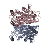
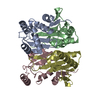
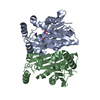
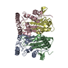
| Title | PRECURSOR OF THE T152C MUTANT GLYCOSYLASPARAGINASE FROM FLAVOBACTERIUM MENINGOSEPTICUM | ||||||
 Components Components | PROTEIN (GLYCOSYLASPARAGINASE) | ||||||
 Keywords Keywords | HYDROLASE/HYDROLASE INHIBITOR / PRECURSOR / GLYCOSYLASPARAGINASE / N-TERMINAL NUCLEOPHILE / AUTOPROTEOLYSIS / MUTANT / HYDROLASE-HYDROLASE INHIBITOR complex | ||||||
| Function / homology |  Function and homology information Function and homology informationN4-(beta-N-acetylglucosaminyl)-L-asparaginase / N4-(beta-N-acetylglucosaminyl)-L-asparaginase activity / peptidase activity / periplasmic space / proteolysis / cytoplasm Similarity search - Function | ||||||
| Biological species |  Elizabethkingia meningoseptica (bacteria) Elizabethkingia meningoseptica (bacteria) | ||||||
| Method |  X-RAY DIFFRACTION / SYNCHROTRON / OTHER / Resolution: 1.9 Å X-RAY DIFFRACTION / SYNCHROTRON / OTHER / Resolution: 1.9 Å | ||||||
 Authors Authors | Guo, H.-C. / Xu, Q. | ||||||
 Citation Citation | Journal: Cell(Cambridge,Mass.) / Year: 1999 Title: Structural insights into the mechanism of intramolecular proteolysis. Authors: Xu, Q. / Buckley, D. / Guan, C. / Guo, H.C. | ||||||
| History |
|
- Structure visualization
| Structure viewer | Molecule: MolmilJmol/JSmol |
|---|
- Downloads & links
-Download
| PDBx/mmCIF format | 9gac.cif.gz | 120.2 KB | Display | PDBx/mmCIF format |
|---|---|---|---|---|
| PDB format | pdb9gac.ent.gz | 91.5 KB | Display | PDB format |
| PDBx/mmJSON format | 9gac.json.gz | Tree view | PDBx/mmJSON format | |
| Others |  Other downloads Other downloads |
-Validation report
| Arichive directory | https://data.pdbj.org/pub/pdb/validation_reports/ga/9gacftp://data.pdbj.org/pub/pdb/validation_reports/ga/9gac | HTTPS FTP |
|---|
-Related structure data
| Related structure data |  9gaaC  9gafC  2gawS S: Starting model for refinement C: citing same article ( |
|---|---|
| Similar structure data |


-Links
 PDBj
PDBj


- Assembly
| Deposited unit | 
| ||||||||
|---|---|---|---|---|---|---|---|---|---|
| 1 |
| ||||||||
| Unit cell |
| ||||||||
| Noncrystallographic symmetry (NCS) | NCS oper: (Code: given Matrix: (-0.99991, -0.01264, 0.00331), Vector: |
-Components
| #1: Protein | Mass: 32181.672 Da / Num. of mol.: 2 / Mutation: T152C Source method: isolated from a genetically manipulated source Details: AN INHIBITOR (FREE GLYCINE) IS BOUND TO EACH PRECURSOR Source: (gene. exp.) Elizabethkingia meningoseptica (bacteria)Description: RECOMBINANT PROTEIN / Plasmid: PMAL FUSION PROTEIN / Production host: References: UniProt: Q47898, N4-(beta-N-acetylglucosaminyl)-L-asparaginase #2: Chemical |   Type: peptide linking / Mass: 75.067 Da / Num. of mol.: 2 / Source method: obtained synthetically / Formula: C2H5NO2 Type: peptide linking / Mass: 75.067 Da / Num. of mol.: 2 / Source method: obtained synthetically / Formula: C2H5NO2#3: Water | ChemComp-HOH / |  Mass: 18.015 Da / Num. of mol.: 151 / Source method: isolated from a natural source / Formula: H2O Mass: 18.015 Da / Num. of mol.: 151 / Source method: isolated from a natural source / Formula: H2ONonpolymer details | A FREE GLYCINE MOLECULE BOUND TO EACH PRECURSOR TO ACT AS THE INHIBITOR OF AUTOPROTEO | |
|---|
-Experimental details
-Experiment
| Experiment | Method: X-RAY DIFFRACTION / Number of used crystals: 1 |
|---|
- Sample preparation
| Crystal | Density Matthews: 2.26 Å3/Da / Density % sol: 45 % | ||||||||||||||||||||||||||||||||||||||||||||||||||||||||||||
|---|---|---|---|---|---|---|---|---|---|---|---|---|---|---|---|---|---|---|---|---|---|---|---|---|---|---|---|---|---|---|---|---|---|---|---|---|---|---|---|---|---|---|---|---|---|---|---|---|---|---|---|---|---|---|---|---|---|---|---|---|---|
| Crystal grow | pH: 7.5 Details: PROTEIN WAS CRYSTALLIZED FROM 15% PEG 3300, 100MM TRIS, PH 7.5, 0.2M LITHIUM SULFATE, 0.1% SODIUM AZIDE | ||||||||||||||||||||||||||||||||||||||||||||||||||||||||||||
| Crystal grow | *PLUS Temperature: 277 K / pH: 7.4 / Method: vapor diffusion, hanging drop | ||||||||||||||||||||||||||||||||||||||||||||||||||||||||||||
| Components of the solutions | *PLUS
|
-Data collection
| Diffraction | Mean temperature: 100 K |
|---|---|
| Diffraction source | Source: SYNCHROTRON / Site: NSLS  / Beamline: X4A / Wavelength: 0.9202 / Beamline: X4A / Wavelength: 0.9202 |
| Detector | Type: RIGAKU RAXIS IV / Detector: IMAGE PLATE / Date: Mar 1, 1998 |
| Radiation | Protocol: SINGLE WAVELENGTH / Monochromatic (M) / Laue (L): M / Scattering type: x-ray |
| Radiation wavelength | Wavelength: 0.9202 Å / Relative weight: 1 |
| Reflection | Resolution: 1.9→25 Å / Num. obs: 41303 / % possible obs: 94.3 % / Observed criterion σ(I): -2 / Redundancy: 2.17 % / Rsym value: 5.6 / Net I/σ(I): 9.4 |
| Reflection shell | Resolution: 1.9→1.97 Å / Redundancy: 1.62 % / Mean I/σ(I) obs: 4 / Rsym value: 18.5 / % possible all: 89.1 |
| Reflection | *PLUS Num. measured all: 89551 / Rmerge(I) obs: 0.056 |
| Reflection shell | *PLUS % possible obs: 89.1 % / Rmerge(I) obs: 0.185 |
- Processing
| Software |
| ||||||||||||||||||||||||||||||||||||||||||||||||||||||||||||||||||||||||||||||||
|---|---|---|---|---|---|---|---|---|---|---|---|---|---|---|---|---|---|---|---|---|---|---|---|---|---|---|---|---|---|---|---|---|---|---|---|---|---|---|---|---|---|---|---|---|---|---|---|---|---|---|---|---|---|---|---|---|---|---|---|---|---|---|---|---|---|---|---|---|---|---|---|---|---|---|---|---|---|---|---|---|---|
| Refinement | Method to determine structure: OTHER Starting model: PDB ENTRY 2GAW Resolution: 1.9→6 Å / Rfactor Rfree error: 0.005 / Data cutoff high absF: 100000 / Isotropic thermal model: RESTRAINED INDIVIDUAL B F / Cross valid method: THROUGHOUT / σ(F): 0
| ||||||||||||||||||||||||||||||||||||||||||||||||||||||||||||||||||||||||||||||||
| Displacement parameters | Biso mean: 10.6 Å2 | ||||||||||||||||||||||||||||||||||||||||||||||||||||||||||||||||||||||||||||||||
| Refine analyze | Luzzati d res low obs: 6 Å | ||||||||||||||||||||||||||||||||||||||||||||||||||||||||||||||||||||||||||||||||
| Refinement step | Cycle: LAST / Resolution: 1.9→6 Å
| ||||||||||||||||||||||||||||||||||||||||||||||||||||||||||||||||||||||||||||||||
| Refine LS restraints |
| ||||||||||||||||||||||||||||||||||||||||||||||||||||||||||||||||||||||||||||||||
| Refine LS restraints NCS | NCS model details: RESTRAINTS | ||||||||||||||||||||||||||||||||||||||||||||||||||||||||||||||||||||||||||||||||
| LS refinement shell | Resolution: 1.9→1.98 Å / Rfactor Rfree error: 0.017 / Total num. of bins used: 8
| ||||||||||||||||||||||||||||||||||||||||||||||||||||||||||||||||||||||||||||||||
| Xplor file |
| ||||||||||||||||||||||||||||||||||||||||||||||||||||||||||||||||||||||||||||||||
| Software | *PLUS Name: X-PLOR / Version: 3.1 / Classification: refinement | ||||||||||||||||||||||||||||||||||||||||||||||||||||||||||||||||||||||||||||||||
| Refine LS restraints | *PLUS
|
