Protocol: SINGLE WAVELENGTH / Monochromatic (M) / Laue (L): M / Scattering type: x-ray
Radiation wavelength
Wavelength: 1.1 Å / Relative weight: 1
Reflection
Resolution: 1.63→61.8 Å / Num. obs: 68901 / % possible obs: 97.3 % / Redundancy: 3.7 % / Net I/σ(I): 0
Reflection shell
Resolution: 1.6→1.641 Å
-
Processing
Software
Name
Version
Classification
REFMAC
5.8.0189
refinement
iMOSFLM
datareduction
Aimless
datascaling
MOLREP
phasing
Refinement
Resolution: 1.63→20 Å / Cor.coef. Fo:Fc: 0.954 / Cor.coef. Fo:Fc free: 0.941 / SU B: 1.347 / SU ML: 0.049 / Cross valid method: THROUGHOUT / ESU R: 0.086 / ESU R Free: 0.086 / Details: HYDROGENS HAVE BEEN ADDED IN THE RIDING POSITIONS
Rfactor
Num. reflection
% reflection
Selection details
Rfree
0.19739
3358
5.1 %
RANDOM
Rwork
0.16873
-
-
-
obs
0.17019
65359
97.03 %
-
Solvent computation
Ion probe radii: 0.8 Å / Shrinkage radii: 0.8 Å / VDW probe radii: 1.2 Å
 Movie
Movie Controller


 Open data
 Basic information
 Components
 Keywords
 Function and homology information
 Elizabethkingia meningoseptica (bacteria)
 X-RAY DIFFRACTION /
  Authors
 United States, 1items
  Citation
 Structure visualization
 Downloads & links
 Other downloads
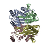


 PDBj
PDBj


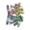
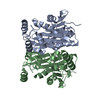
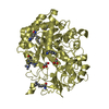
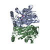
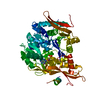
 Assembly
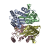
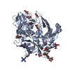
 Mass: 18.015 Da / Num. of mol.: 222 / Source method: isolated from a natural source / Formula: H2O
 Sample preparation
 Processing