 Movie
Movie Controller


+ Open data

- Basic information
| Entry | Database: PDB / ID: 9g99 | |||||||||
|---|---|---|---|---|---|---|---|---|---|---|
| Title | Joint neutron and x-ray structure of apo alginate lyase PsAlg7C | |||||||||
 Components Components | Alginate lyase | |||||||||
 Keywords Keywords | LYASE / complex / beta jellyroll / alginate | |||||||||
| Function / homology | Alginate lyase 2 / Alginate lyase / Concanavalin A-like lectin/glucanase domain superfamily / lyase activity / Alginate lyase Function and homology information Function and homology information | |||||||||
| Biological species |  Paradendryphiella salina (fungus) Paradendryphiella salina (fungus) | |||||||||
| Method |  X-RAY DIFFRACTION / NEUTRON DIFFRACTION / MOLECULAR REPLACEMENT / Resolution: 2.15 Å X-RAY DIFFRACTION / NEUTRON DIFFRACTION / MOLECULAR REPLACEMENT / Resolution: 2.15 Å | |||||||||
 Authors Authors | Wilkens, C. / Meilleur, F. / Morth, J.P. | |||||||||
| Funding support |  Denmark, 1items Denmark, 1items
| |||||||||
 Citation Citation | Journal: Nat Commun / Year: 2025 Title: Unraveling the molecular mechanism of polysaccharide lyases for efficient alginate degradation. Authors: Rivas-Fernandez, J.P. / Vuillemin, M. / Pilgaard, B. / Klau, L.J. / Fredslund, F. / Lund-Hanssen, C. / Welner, D.H. / Meyer, A.S. / Morth, J.P. / Meilleur, F. / Aachmann, F.L. / Rovira, C. / Wilkens, C. | |||||||||
| History |
|
- Structure visualization
| Structure viewer | Molecule: MolmilJmol/JSmol |
|---|
- Downloads & links
-Download
| PDBx/mmCIF format | 9g99.cif.gz | 126.2 KB | Display | PDBx/mmCIF format |
|---|---|---|---|---|
| PDB format | pdb9g99.ent.gz | 90.9 KB | Display | PDB format |
| PDBx/mmJSON format | 9g99.json.gz | Tree view | PDBx/mmJSON format | |
| Others |  Other downloads Other downloads |
-Validation report
| Summary document | 9g99_validation.pdf.gz | 374.2 KB | Display | wwPDB validaton report |
|---|---|---|---|---|
| Full document | 9g99_full_validation.pdf.gz | 374.1 KB | Display | |
| Data in XML | 9g99_validation.xml.gz | 6.7 KB | Display | |
| Data in CIF | 9g99_validation.cif.gz | 12.1 KB | Display | |
| Arichive directory | https://data.pdbj.org/pub/pdb/validation_reports/g9/9g99ftp://data.pdbj.org/pub/pdb/validation_reports/g9/9g99 | HTTPS FTP |
-Related structure data
| Related structure data | 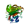 6ywfC  7nczC  7nl3C  7nm6C  7nppC 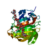 7ny3C  7o6hC  7oofC  7oryC  7p25C  7p90C  7pbfC 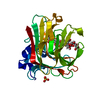 8bjoC  8bxzC  8c0mC  8c3xC  8p6oC  8pc3C  8pc8C  8pcxC  8pdtC  8pedC  8qizC  8qliC 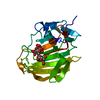 8qmjC  8r43C  8rbiC  9g98C C: citing same article ( |
|---|---|
| Similar structure data |
-Links
 PDBj
PDBj- Assembly
| Deposited unit | 
| ||||||||||||
|---|---|---|---|---|---|---|---|---|---|---|---|---|---|
| 1 |
| ||||||||||||
| Unit cell |
|
-Components
| #1: Protein | Mass: 26713.744 Da / Num. of mol.: 1 Source method: isolated from a genetically manipulated source Source: (gene. exp.) Paradendryphiella salina (fungus) / Gene: PsAlg7C / Production host: Komagataella pastoris (fungus) / References: UniProt: A0A7I9C8Z1 |
|---|---|
| #2: Water | ChemComp-HOH /  Mass: 18.015 Da / Num. of mol.: 199 / Source method: isolated from a natural source / Formula: H2O Mass: 18.015 Da / Num. of mol.: 199 / Source method: isolated from a natural source / Formula: H2O |
| Has protein modification | N |
-Experimental details
-Experiment
| Experiment |
|
|---|
- Sample preparation
| Crystal | Density Matthews: 2.27 Å3/Da / Density % sol: 45.7 % |
|---|---|
| Crystal grow | Temperature: 293 K / Method: vapor diffusion, sitting drop / Details: 30% PEG3350,0.1M Bis-Tris pH 5.5, 0.3M NaCl |
-Data collection
| Diffraction |
| |||||||||||||||||||||||||||
|---|---|---|---|---|---|---|---|---|---|---|---|---|---|---|---|---|---|---|---|---|---|---|---|---|---|---|---|---|
| Diffraction source |
| |||||||||||||||||||||||||||
| Detector |
| |||||||||||||||||||||||||||
| Radiation |
| |||||||||||||||||||||||||||
| Radiation wavelength |
| |||||||||||||||||||||||||||
| Reflection | Entry-ID: 9G99
| |||||||||||||||||||||||||||
| Reflection shell | Rpim(I) all: 0.14
|

- Processing
| Refinement | Biso mean: 20.04 Å2 / Cross valid method: FREE R-VALUE / Method to determine structure:
| |||||||||||||||||||||||||||||||||
|---|---|---|---|---|---|---|---|---|---|---|---|---|---|---|---|---|---|---|---|---|---|---|---|---|---|---|---|---|---|---|---|---|---|---|
| Refinement step | Cycle: LAST / Resolution: 2.15→13.85 Å
| |||||||||||||||||||||||||||||||||
| Refine LS restraints | Type: f_dihedral_angle_d / Dev ideal: 18.185 / Number: 1086 | |||||||||||||||||||||||||||||||||
| LS refinement shell | Resolution: 4.43→27.23 Å
|
