 Movie
Movie Controller

[English] 日本語
 Yorodumi
- PDB-9f8l: Crystal Structure of PhzA/B from Burkholderia cepacia R18194 in c... -
+ Open data

- Basic information
| Entry | Database: PDB / ID: 9f8l | ||||||
|---|---|---|---|---|---|---|---|
| Title | Crystal Structure of PhzA/B from Burkholderia cepacia R18194 in complex with [6-Hydroxy-2-(4-hydroxyphenyl)benzo[b]thiophen-3-yl](3-hydroxyphenyl)methanone | ||||||
 Components Components | Phenazine biosynthesis protein A/B | ||||||
 Keywords Keywords | BIOSYNTHETIC PROTEIN / pyocyanin / phenazine biosynthesis / virulence / inhibitor / ketosteroid-isomerase / cocrystal / PhzA/B / Burkholderia cepacia | ||||||
| Function / homology | Phenazine biosynthesis protein A/B / Phenazine biosynthesis protein A/B / antibiotic biosynthetic process / NTF2-like domain superfamily / : / Phenazine biosynthesis protein A/B Function and homology information Function and homology information | ||||||
| Biological species |  Burkholderia lata (bacteria) Burkholderia lata (bacteria) | ||||||
| Method |  X-RAY DIFFRACTION / SYNCHROTRON / MOLECULAR REPLACEMENT / Resolution: 1.38 Å X-RAY DIFFRACTION / SYNCHROTRON / MOLECULAR REPLACEMENT / Resolution: 1.38 Å | ||||||
 Authors Authors | Thiemann, M. / Zimmermann, M. / Kunick, C. / Blankenfeldt, W. | ||||||
| Funding support | 1items
| ||||||
 Citation Citation | Journal: J.Med.Chem. / Year: 2025 Title: From Bones to Bugs: Structure-Based Development of Raloxifene-Derived Pathoblockers That Inhibit Pyocyanin Production in Pseudomonas aeruginosa. Authors: Thiemann, M. / Zimmermann, M. / Diederich, C. / Zhan, H. / Lebedev, M. / Pletz, J. / Baumgarten, J. / Handke, M. / Musken, M. / Breinbauer, R. / Krasteva-Christ, G. / Zanin, E. / Empting, M. ...Authors: Thiemann, M. / Zimmermann, M. / Diederich, C. / Zhan, H. / Lebedev, M. / Pletz, J. / Baumgarten, J. / Handke, M. / Musken, M. / Breinbauer, R. / Krasteva-Christ, G. / Zanin, E. / Empting, M. / Schiedel, M. / Kunick, C. / Blankenfeldt, W. | ||||||
| History |
|
- Structure visualization
| Structure viewer | Molecule: MolmilJmol/JSmol |
|---|
- Downloads & links
-Download
| PDBx/mmCIF format | 9f8l.cif.gz | 214.8 KB | Display | PDBx/mmCIF format |
|---|---|---|---|---|
| PDB format | pdb9f8l.ent.gz | Display | PDB format | |
| PDBx/mmJSON format | 9f8l.json.gz | Tree view | PDBx/mmJSON format | |
| Others |  Other downloads Other downloads |
-Validation report
| Arichive directory | https://data.pdbj.org/pub/pdb/validation_reports/f8/9f8lftp://data.pdbj.org/pub/pdb/validation_reports/f8/9f8l | HTTPS FTP |
|---|
-Related structure data
| Related structure data |  9f8hC  9f8iC  9f8jC  9f8kC  9f8mC  9f8nC  9f8oC 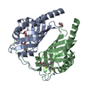 9f8pC  9f8qC 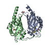 9f8rC  9f8sC C: citing same article ( |
|---|---|
| Similar structure data |
-Links
 PDBj
PDBj
- Assembly
| Deposited unit | 
| ||||||||||||
|---|---|---|---|---|---|---|---|---|---|---|---|---|---|
| 1 | 
| ||||||||||||
| 2 | 
| ||||||||||||
| Unit cell |
| ||||||||||||
| Components on special symmetry positions |
|
-Components
| #1: Protein | Mass: 21531.025 Da / Num. of mol.: 2 Source method: isolated from a genetically manipulated source Source: (gene. exp.) Burkholderia lata (bacteria) / Gene: Bcep18194_B1568 / Plasmid: pET15b / Production host: #2: Chemical |   Mass: 195.237 Da / Num. of mol.: 2 / Source method: obtained synthetically / Formula: C6H13NO4S / Comment: pH buffer*YM Mass: 195.237 Da / Num. of mol.: 2 / Source method: obtained synthetically / Formula: C6H13NO4S / Comment: pH buffer*YM#3: Chemical | Mass: 362.398 Da / Num. of mol.: 2 / Source method: obtained synthetically / Formula: C21H14O4S / Feature type: SUBJECT OF INVESTIGATION #4: Chemical | ChemComp-GOL /   Mass: 92.094 Da / Num. of mol.: 4 / Source method: obtained synthetically / Formula: C3H8O3 Mass: 92.094 Da / Num. of mol.: 4 / Source method: obtained synthetically / Formula: C3H8O3#5: Water | ChemComp-HOH / |  Mass: 18.015 Da / Num. of mol.: 346 / Source method: isolated from a natural source / Formula: H2O Mass: 18.015 Da / Num. of mol.: 346 / Source method: isolated from a natural source / Formula: H2OHas ligand of interest | Y | Has protein modification | N | |
|---|
-Experimental details
-Experiment
| Experiment | Method: X-RAY DIFFRACTION / Number of used crystals: 1 |
|---|
- Sample preparation
| Crystal | Density Matthews: 2.37 Å3/Da / Density % sol: 48.2 % |
|---|---|
| Crystal grow | Temperature: 293 K / Method: vapor diffusion, sitting drop Details: 15% (w/v) PEG monomethyl ether, 0.1 M MES pH 6.14, 0.122 M ammonium acetate, 5% (v/v) glycerol |
-Data collection
| Diffraction | Mean temperature: 100 K / Serial crystal experiment: N |
|---|---|
| Diffraction source | Source: SYNCHROTRON / Site: PETRA III, DESY  / Beamline: P11 / Wavelength: 1.0332 Å / Beamline: P11 / Wavelength: 1.0332 Å |
| Detector | Type: DECTRIS EIGER2 X 16M / Detector: PIXEL / Date: May 19, 2022 |
| Radiation | Protocol: SINGLE WAVELENGTH / Monochromatic (M) / Laue (L): M / Scattering type: x-ray |
| Radiation wavelength | Wavelength: 1.0332 Å / Relative weight: 1 |
| Reflection | Resolution: 1.38→53.18 Å / Num. obs: 79713 / % possible obs: 99.1 % / Redundancy: 6.9 % / CC1/2: 0.999 / Rmerge(I) obs: 0.047 / Rpim(I) all: 0.019 / Rrim(I) all: 0.051 / Net I/σ(I): 16.5 |
| Reflection shell | Resolution: 1.38→1.4 Å / Redundancy: 6.9 % / Rmerge(I) obs: 0.686 / Mean I/σ(I) obs: 2.1 / Num. unique obs: 3936 / CC1/2: 0.897 / Rpim(I) all: 0.278 / Rrim(I) all: 0.741 |
- Processing
| Software |
| |||||||||||||||||||||||||||||||||||||||||||||||||||||||||||||||||||||||||||||||||||||||||||||||||||||||||||||||||||||||||||||||||||||||||||||||||||||||||||||||||||||||||||||||||||||||||||||||||||||||||||
|---|---|---|---|---|---|---|---|---|---|---|---|---|---|---|---|---|---|---|---|---|---|---|---|---|---|---|---|---|---|---|---|---|---|---|---|---|---|---|---|---|---|---|---|---|---|---|---|---|---|---|---|---|---|---|---|---|---|---|---|---|---|---|---|---|---|---|---|---|---|---|---|---|---|---|---|---|---|---|---|---|---|---|---|---|---|---|---|---|---|---|---|---|---|---|---|---|---|---|---|---|---|---|---|---|---|---|---|---|---|---|---|---|---|---|---|---|---|---|---|---|---|---|---|---|---|---|---|---|---|---|---|---|---|---|---|---|---|---|---|---|---|---|---|---|---|---|---|---|---|---|---|---|---|---|---|---|---|---|---|---|---|---|---|---|---|---|---|---|---|---|---|---|---|---|---|---|---|---|---|---|---|---|---|---|---|---|---|---|---|---|---|---|---|---|---|---|---|---|---|---|---|---|---|---|
| Refinement | Method to determine structure: MOLECULAR REPLACEMENT / Resolution: 1.38→42.99 Å / SU ML: 0.14 / Cross valid method: THROUGHOUT / σ(F): 1.36 / Phase error: 16.79 / Stereochemistry target values: ML
| |||||||||||||||||||||||||||||||||||||||||||||||||||||||||||||||||||||||||||||||||||||||||||||||||||||||||||||||||||||||||||||||||||||||||||||||||||||||||||||||||||||||||||||||||||||||||||||||||||||||||||
| Solvent computation | Shrinkage radii: 0.9 Å / VDW probe radii: 1.1 Å / Solvent model: FLAT BULK SOLVENT MODEL | |||||||||||||||||||||||||||||||||||||||||||||||||||||||||||||||||||||||||||||||||||||||||||||||||||||||||||||||||||||||||||||||||||||||||||||||||||||||||||||||||||||||||||||||||||||||||||||||||||||||||||
| Displacement parameters | Biso mean: 25.879 Å2 | |||||||||||||||||||||||||||||||||||||||||||||||||||||||||||||||||||||||||||||||||||||||||||||||||||||||||||||||||||||||||||||||||||||||||||||||||||||||||||||||||||||||||||||||||||||||||||||||||||||||||||
| Refinement step | Cycle: LAST / Resolution: 1.38→42.99 Å
| |||||||||||||||||||||||||||||||||||||||||||||||||||||||||||||||||||||||||||||||||||||||||||||||||||||||||||||||||||||||||||||||||||||||||||||||||||||||||||||||||||||||||||||||||||||||||||||||||||||||||||
| Refine LS restraints |
| |||||||||||||||||||||||||||||||||||||||||||||||||||||||||||||||||||||||||||||||||||||||||||||||||||||||||||||||||||||||||||||||||||||||||||||||||||||||||||||||||||||||||||||||||||||||||||||||||||||||||||
| LS refinement shell |
|
