 Movie
Movie Controller

[English] 日本語
 Yorodumi
- PDB-9euk: Cryo-EM structure of Staphylococcus aureus bacteriophage phi812 b... -
+ Open data

- Basic information
| Entry | Database: PDB / ID: 9euk | |||||||||||||||||||||||||||
|---|---|---|---|---|---|---|---|---|---|---|---|---|---|---|---|---|---|---|---|---|---|---|---|---|---|---|---|---|
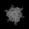
| Title | Cryo-EM structure of Staphylococcus aureus bacteriophage phi812 baseplate in the post-contraction state - sheath initiator, wedge module, and inner tripod proteins | |||||||||||||||||||||||||||
 Components Components |
| |||||||||||||||||||||||||||
 Keywords Keywords | VIRUS / bacteriophage / phage / contractile / phi812 / baseplate | |||||||||||||||||||||||||||
| Function / homology |  Function and homology information Function and homology informationDomain of unknown function DUF4815 / Domain of unknown function (DUF4815) / Protein of unknown function DUF2634 / Contractile injection system sheath initiator / Baseplate protein J-like / Baseplate J-like protein barrel domain / LysM domain superfamily / LysM domain Similarity search - Domain/homology | |||||||||||||||||||||||||||
| Biological species |  Staphylococcus phage 812 (virus) Staphylococcus phage 812 (virus) | |||||||||||||||||||||||||||
| Method | ELECTRON MICROSCOPY / single particle reconstruction / cryo EM / Resolution: 3.1 Å | |||||||||||||||||||||||||||
 Authors Authors | Binovsky, J. / Siborova, M. / Baska, R. / Pichel-Beleiro, A. / Skubnik, K. / Novacek, J. / van Raaij, M.J. / Plevka, P. | |||||||||||||||||||||||||||
| Funding support | European Union,  Czech Republic, 2items Czech Republic, 2items
| |||||||||||||||||||||||||||
 Citation Citation | Journal: To Be Published Title: Cell attachment and tail contraction of S. aureus phage phi812 Authors: Binovsky, J. / Siborova, M. / Baska, R. / Pichel-Beleiro, A. / Skubnik, K. / Novacek, J. / van Raaij, M.J. / Plevka, P. | |||||||||||||||||||||||||||
| History |
|
- Structure visualization
| Structure viewer | Molecule: MolmilJmol/JSmol |
|---|
- Downloads & links
-Download
| PDBx/mmCIF format | 9euk.cif.gz | 733.3 KB | Display | PDBx/mmCIF format |
|---|---|---|---|---|
| PDB format | pdb9euk.ent.gz | 583.3 KB | Display | PDB format |
| PDBx/mmJSON format | 9euk.json.gz | Tree view | PDBx/mmJSON format | |
| Others |  Other downloads Other downloads |
-Validation report
| Arichive directory | https://data.pdbj.org/pub/pdb/validation_reports/eu/9eukftp://data.pdbj.org/pub/pdb/validation_reports/eu/9euk | HTTPS FTP |
|---|
-Related structure data
| Related structure data |  19973MC  9eufC  9eugC  9euhC 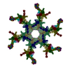 9euiC 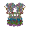 9eujC 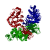 9eulC  9eumC  9f04C  9f05C  9f06C  9fkoC M: map data used to model this data C: citing same article ( |
|---|---|
| Similar structure data |


-Links
 PDBj
PDBj
- Assembly
| Deposited unit | 
|
|---|---|
| 1 | 
|
-Components
| #1: Protein | Mass: 26611.752 Da / Num. of mol.: 1 / Source method: isolated from a natural source / Source: (natural) Staphylococcus phage 812 (virus) / References: UniProt: A0A0U1UXD7 | ||||||
|---|---|---|---|---|---|---|---|
| #2: Protein | Mass: 39248.859 Da / Num. of mol.: 2 / Source method: isolated from a natural source / Source: (natural) Staphylococcus phage 812 (virus) / References: UniProt: A0A0U1WF63#3: Protein | | Mass: 116391.883 Da / Num. of mol.: 1 / Source method: isolated from a natural source / Source: (natural) Staphylococcus phage 812 (virus) / References: UniProt: A0A0U1WGD3#4: Protein | Mass: 129262.961 Da / Num. of mol.: 3 / Source method: isolated from a natural source / Source: (natural) Staphylococcus phage 812 (virus) / References: UniProt: A0A8E5NSA0Has protein modification | N | |
-Experimental details
-Experiment
| Experiment | Method: ELECTRON MICROSCOPY |
|---|---|
| EM experiment | Aggregation state: PARTICLE / 3D reconstruction method: single particle reconstruction |
- Sample preparation
| Component |
| ||||||||||||||||||||||||
|---|---|---|---|---|---|---|---|---|---|---|---|---|---|---|---|---|---|---|---|---|---|---|---|---|---|
| Source (natural) |
| ||||||||||||||||||||||||
| Details of virus | Empty: YES / Enveloped: NO / Isolate: SPECIES / Type: VIRION | ||||||||||||||||||||||||
| Buffer solution | pH: 8 / Details: 50mM Tris, 10mM NaCl, 10mM CaCl2 | ||||||||||||||||||||||||
| Specimen | Embedding applied: NO / Shadowing applied: NO / Staining applied: NO / Vitrification applied: YES | ||||||||||||||||||||||||
| Specimen support | Grid material: COPPER / Grid mesh size: 200 divisions/in. / Grid type: Quantifoil R2/1 | ||||||||||||||||||||||||
| Vitrification | Instrument: FEI VITROBOT MARK IV / Cryogen name: ETHANE / Humidity: 100 % / Chamber temperature: 277 K |
- Electron microscopy imaging
| Experimental equipment |  Model: Titan Krios / Image courtesy: FEI Company |
|---|---|
| Microscopy | Model: FEI TITAN KRIOS |
| Electron gun | Electron source:  FIELD EMISSION GUN / Accelerating voltage: 300 kV / Illumination mode: FLOOD BEAM FIELD EMISSION GUN / Accelerating voltage: 300 kV / Illumination mode: FLOOD BEAM |
| Electron lens | Mode: BRIGHT FIELD / Nominal magnification: 130000 X / Nominal defocus max: 3000 nm / Nominal defocus min: 800 nm / Cs: 2.7 mm / C2 aperture diameter: 70 µm |
| Image recording | Average exposure time: 7 sec. / Electron dose: 42 e/Å2 / Detector mode: COUNTING / Film or detector model: GATAN K2 SUMMIT (4k x 4k) / Num. of real images: 15371 |
| EM imaging optics | Energyfilter name: GIF Quantum LS / Energyfilter slit width: 20 eV |
| Image scans | Width: 3838 / Height: 3710 / Movie frames/image: 40 / Used frames/image: 1-40 |
- Processing
| EM software |
| ||||||||||||||||||||||||||||||||||||
|---|---|---|---|---|---|---|---|---|---|---|---|---|---|---|---|---|---|---|---|---|---|---|---|---|---|---|---|---|---|---|---|---|---|---|---|---|---|
| CTF correction | Type: PHASE FLIPPING AND AMPLITUDE CORRECTION | ||||||||||||||||||||||||||||||||||||
| Particle selection | Num. of particles selected: 54841 | ||||||||||||||||||||||||||||||||||||
| Symmetry | Point symmetry: C6 (6 fold cyclic) | ||||||||||||||||||||||||||||||||||||
| 3D reconstruction | Resolution: 3.1 Å / Resolution method: FSC 0.143 CUT-OFF / Num. of particles: 25203 / Symmetry type: POINT | ||||||||||||||||||||||||||||||||||||
| Atomic model building | Protocol: FLEXIBLE FIT / Space: REAL | ||||||||||||||||||||||||||||||||||||
| Refine LS restraints |
|
