 Movie
Movie Controller


+ Open data

- Basic information
| Entry | Database: PDB / ID: 9eoh | |||||||||
|---|---|---|---|---|---|---|---|---|---|---|
| Title | PHF type tau filament from in vitro V337M mutant | |||||||||
 Components Components | Isoform Tau-F of Microtubule-associated protein tau | |||||||||
 Keywords Keywords | PROTEIN FIBRIL / PHF / tau filament / V337M | |||||||||
| Function / homology |  Function and homology information Function and homology informationplus-end-directed organelle transport along microtubule / histone-dependent DNA binding / negative regulation of protein localization to mitochondrion / neurofibrillary tangle / microtubule lateral binding / axonal transport / tubulin complex / positive regulation of protein localization to synapse / phosphatidylinositol bisphosphate binding / generation of neurons ...plus-end-directed organelle transport along microtubule / histone-dependent DNA binding / negative regulation of protein localization to mitochondrion / neurofibrillary tangle / microtubule lateral binding / axonal transport / tubulin complex / positive regulation of protein localization to synapse / phosphatidylinositol bisphosphate binding / generation of neurons / rRNA metabolic process / axonal transport of mitochondrion / regulation of mitochondrial fission / axon development / regulation of microtubule-based movement / intracellular distribution of mitochondria / regulation of chromosome organization / central nervous system neuron development / minor groove of adenine-thymine-rich DNA binding / lipoprotein particle binding / microtubule polymerization / negative regulation of mitochondrial membrane potential / regulation of microtubule polymerization / dynactin binding / apolipoprotein binding / protein polymerization / main axon / Caspase-mediated cleavage of cytoskeletal proteins / regulation of microtubule polymerization or depolymerization / negative regulation of mitochondrial fission / axolemma / glial cell projection / neurofibrillary tangle assembly / positive regulation of axon extension / regulation of cellular response to heat / positive regulation of microtubule polymerization / positive regulation of protein localization / Activation of AMPK downstream of NMDARs / positive regulation of superoxide anion generation / cellular response to brain-derived neurotrophic factor stimulus / regulation of long-term synaptic depression / supramolecular fiber organization / cytoplasmic microtubule organization / regulation of calcium-mediated signaling / axon cytoplasm / somatodendritic compartment / synapse assembly / phosphatidylinositol binding / nuclear periphery / astrocyte activation / enzyme inhibitor activity / protein phosphatase 2A binding / stress granule assembly / regulation of microtubule cytoskeleton organization / regulation of autophagy / cellular response to reactive oxygen species / microglial cell activation / cellular response to nerve growth factor stimulus / Hsp90 protein binding / protein homooligomerization / SH3 domain binding / PKR-mediated signaling / synapse organization / regulation of synaptic plasticity / response to lead ion / microtubule cytoskeleton organization / memory / neuron projection development / cytoplasmic ribonucleoprotein granule / cell-cell signaling / cellular response to heat / single-stranded DNA binding / growth cone / protein-folding chaperone binding / microtubule cytoskeleton / actin binding / cell body / double-stranded DNA binding / sequence-specific DNA binding / microtubule binding / amyloid fibril formation / dendritic spine / microtubule / learning or memory / protein-macromolecule adaptor activity / neuron projection / membrane raft / negative regulation of gene expression / axon / neuronal cell body / DNA damage response / dendrite / protein kinase binding / enzyme binding / mitochondrion / DNA binding / RNA binding / extracellular region / identical protein binding / nucleus Similarity search - Function | |||||||||
| Biological species |  Homo sapiens (human) Homo sapiens (human) | |||||||||
| Method | ELECTRON MICROSCOPY / helical reconstruction / cryo EM / Resolution: 2.8 Å | |||||||||
 Authors Authors | Qi, C. / Lovestam, S. / Scheres, S.H.W. / Michel, G. | |||||||||
| Funding support |  United Kingdom, 2items United Kingdom, 2items
| |||||||||
 Citation Citation | Journal: Nat Struct Mol Biol / Year: 2025 Title: Tau filaments with the Alzheimer fold in human MAPT mutants V337M and R406W. Authors: Chao Qi / Sofia Lövestam / Alexey G Murzin / Sew Peak-Chew / Catarina Franco / Marika Bogdani / Caitlin Latimer / Jill R Murrell / Patrick W Cullinane / Zane Jaunmuktane / Thomas D Bird / ...Authors: Chao Qi / Sofia Lövestam / Alexey G Murzin / Sew Peak-Chew / Catarina Franco / Marika Bogdani / Caitlin Latimer / Jill R Murrell / Patrick W Cullinane / Zane Jaunmuktane / Thomas D Bird / Bernardino Ghetti / Sjors H W Scheres / Michel Goedert /  Abstract: Frontotemporal dementia (FTD) and Alzheimer's disease (AD) are the most common forms of early-onset dementia. Unlike AD, FTD begins with behavioral changes before the development of cognitive ...Frontotemporal dementia (FTD) and Alzheimer's disease (AD) are the most common forms of early-onset dementia. Unlike AD, FTD begins with behavioral changes before the development of cognitive impairment. Dominantly inherited mutations in MAPT, the microtubule-associated protein tau gene, give rise to cases of FTD and parkinsonism linked to chromosome 17. These individuals develop abundant filamentous tau inclusions in brain cells in the absence of β-amyloid deposits. Here, we used cryo-electron microscopy to determine the structures of tau filaments from the brains of human MAPT mutants V337M and R406W. Both amino acid substitutions gave rise to tau filaments with the Alzheimer fold, which consisted of paired helical filaments in all V337M and R406W cases and of straight filaments in two V337M cases. We also identified another assembly of the Alzheimer fold into triple tau filaments in a V337M case. Filaments assembled from recombinant tau (297-391) with substitution V337M had the Alzheimer fold and showed an increased rate of assembly. #1: Journal: bioRxiv / Year: 2024 Title: Tau filaments with the Alzheimer fold in cases with mutations V337M and R406W. Authors: Chao Qi / Sofia Lövestam / Alexey G Murzin / Sew Peak-Chew / Catarina Franco / Marika Bogdani / Caitlin Latimer / Jill R Murrell / Patrick W Cullinane / Zane Jaunmuktane / Thomas D Bird / ...Authors: Chao Qi / Sofia Lövestam / Alexey G Murzin / Sew Peak-Chew / Catarina Franco / Marika Bogdani / Caitlin Latimer / Jill R Murrell / Patrick W Cullinane / Zane Jaunmuktane / Thomas D Bird / Bernardino Ghetti / Sjors H W Scheres / Michel Goedert / Abstract: Frontotemporal dementia (FTD) and Alzheimer's disease are the most common forms of early-onset dementia. Dominantly inherited mutations in , the microtubule-associated protein tau gene, cause FTD and ...Frontotemporal dementia (FTD) and Alzheimer's disease are the most common forms of early-onset dementia. Dominantly inherited mutations in , the microtubule-associated protein tau gene, cause FTD and parkinsonism linked to chromosome 17 (FTDP-17). Individuals with FTDP-17 develop abundant filamentous tau inclusions in brain cells. Here we used electron cryo-microscopy to determine the structures of tau filaments from the brains of individuals with mutations V337M and R406W. Both mutations gave rise to tau filaments with the Alzheimer fold, which consisted of paired helical filaments in all V337M and R406W cases and of straight filaments in two V337M cases. We also identified a new assembly of the Alzheimer fold into triple tau filaments in a V337M case. Filaments assembled from recombinant tau(297-391) with mutation V337M had the Alzheimer fold and showed an increased rate of assembly. | |||||||||
| History |
|
- Structure visualization
| Structure viewer | Molecule: MolmilJmol/JSmol |
|---|
- Downloads & links
-Download
| PDBx/mmCIF format | 9eoh.cif.gz | 121.9 KB | Display | PDBx/mmCIF format |
|---|---|---|---|---|
| PDB format | pdb9eoh.ent.gz | 80.3 KB | Display | PDB format |
| PDBx/mmJSON format | 9eoh.json.gz | Tree view | PDBx/mmJSON format | |
| Others |  Other downloads Other downloads |
-Validation report
| Arichive directory | https://data.pdbj.org/pub/pdb/validation_reports/eo/9eohftp://data.pdbj.org/pub/pdb/validation_reports/eo/9eoh | HTTPS FTP |
|---|
-Related structure data
| Related structure data |  19855MC 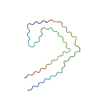 9eo7C 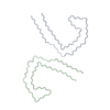 9eo9C 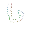 9eoeC  9eogC C: citing same article ( M: map data used to model this data |
|---|---|
| Similar structure data |


-Links
 PDBj
PDBj


- Assembly
| Deposited unit | 
|
|---|---|
| 1 |
|
-Components
| #1: Protein | Mass: 45951.938 Da / Num. of mol.: 6 Source method: isolated from a genetically manipulated source Source: (gene. exp.) Homo sapiens (human) / Gene: MAPT, MAPTL, MTBT1, TAU / Production host:  Has protein modification | N | |
|---|
-Experimental details
-Experiment
| Experiment | Method: ELECTRON MICROSCOPY |
|---|---|
| EM experiment | Aggregation state: FILAMENT / 3D reconstruction method: helical reconstruction |
- Sample preparation
| Component | Name: PHF tau filament from in vitro V337M / Type: COMPLEX / Entity ID: all / Source: RECOMBINANT |
|---|---|
| Source (natural) | Organism: Homo sapiens (human) |
| Source (recombinant) | Organism: |
| Buffer solution | pH: 8 |
| Specimen | Embedding applied: NO / Shadowing applied: NO / Staining applied: NO / Vitrification applied: YES |
| Vitrification | Cryogen name: ETHANE |
- Electron microscopy imaging
| Experimental equipment |  Model: Titan Krios / Image courtesy: FEI Company |
|---|---|
| Microscopy | Model: FEI TITAN KRIOS |
| Electron gun | Electron source:  FIELD EMISSION GUN / Accelerating voltage: 300 kV / Illumination mode: FLOOD BEAM FIELD EMISSION GUN / Accelerating voltage: 300 kV / Illumination mode: FLOOD BEAM |
| Electron lens | Mode: BRIGHT FIELD / Nominal defocus max: 2000 nm / Nominal defocus min: 1000 nm |
| Image recording | Electron dose: 40 e/Å2 / Film or detector model: FEI FALCON IV (4k x 4k) |
- Processing
| CTF correction | Type: NONE |
|---|---|
| Helical symmerty | Angular rotation/subunit: 179.2 ° / Axial rise/subunit: 2.37 Å / Axial symmetry: C1 |
| 3D reconstruction | Resolution: 2.8 Å / Resolution method: FSC 0.143 CUT-OFF / Num. of particles: 37720 / Symmetry type: HELICAL |
