- EMDB-19854: PHF type tau filament from R406W mutant -
+
Open data
ID or keywords:
Loading...
-
Basic information
Entry
Database: EMDB / ID: EMD-19854
Title
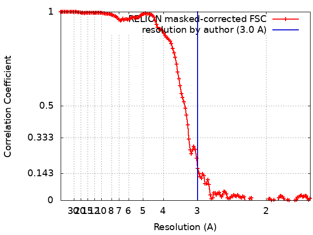
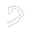
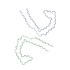
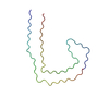
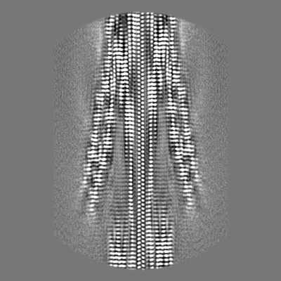
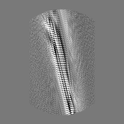
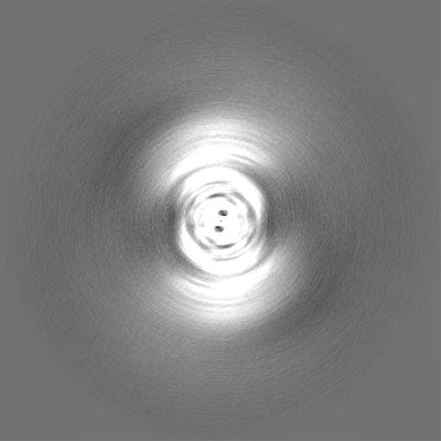
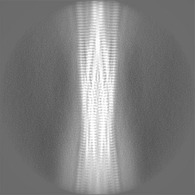
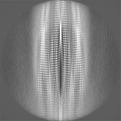
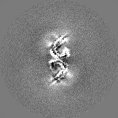
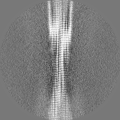
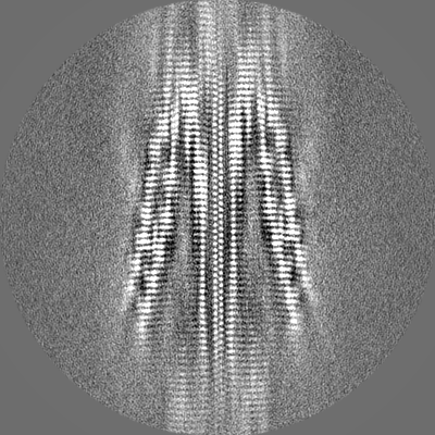
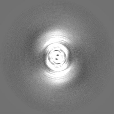
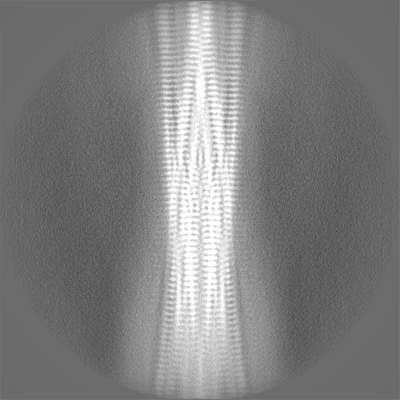
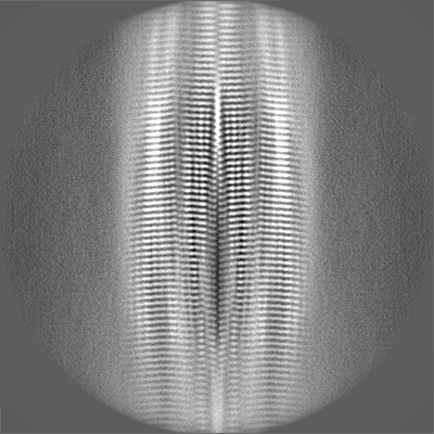
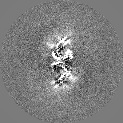
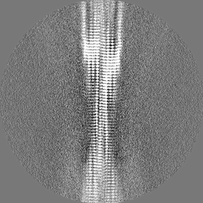
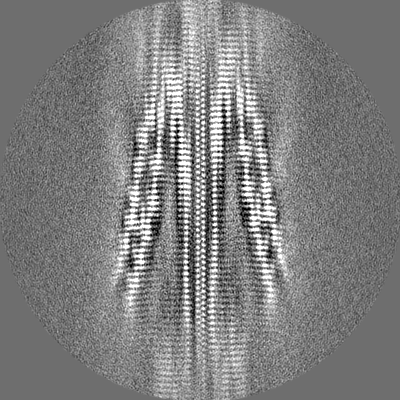
PHF type tau filament from R406W mutant
Map data
Sample
Complex: PHF tau filament from mutant R406W
Protein or peptide: Microtubule-associated protein tau
Keywords
TF / tau filament / R406W / PROTEIN FIBRIL
Function / homology
Function and homology information
plus-end-directed organelle transport along microtubule / histone-dependent DNA binding / negative regulation of protein localization to mitochondrion / neurofibrillary tangle / microtubule lateral binding / axonal transport / tubulin complex / positive regulation of protein localization to synapse / phosphatidylinositol bisphosphate binding / generation of neurons ...plus-end-directed organelle transport along microtubule / histone-dependent DNA binding / negative regulation of protein localization to mitochondrion / neurofibrillary tangle / microtubule lateral binding / axonal transport / tubulin complex / positive regulation of protein localization to synapse / phosphatidylinositol bisphosphate binding / generation of neurons / rRNA metabolic process / axonal transport of mitochondrion / axon development / regulation of mitochondrial fission / regulation of microtubule-based movement / intracellular distribution of mitochondria / regulation of chromosome organization / central nervous system neuron development / minor groove of adenine-thymine-rich DNA binding / lipoprotein particle binding / microtubule polymerization / negative regulation of mitochondrial membrane potential / regulation of microtubule polymerization / dynactin binding / apolipoprotein binding / protein polymerization / main axon / Caspase-mediated cleavage of cytoskeletal proteins / regulation of microtubule polymerization or depolymerization / negative regulation of mitochondrial fission / axolemma / glial cell projection / neurofibrillary tangle assembly / positive regulation of axon extension / regulation of cellular response to heat / positive regulation of microtubule polymerization / positive regulation of protein localization / Activation of AMPK downstream of NMDARs / positive regulation of superoxide anion generation / cellular response to brain-derived neurotrophic factor stimulus / regulation of long-term synaptic depression / supramolecular fiber organization / cytoplasmic microtubule organization / regulation of calcium-mediated signaling / axon cytoplasm / somatodendritic compartment / synapse assembly / phosphatidylinositol binding / nuclear periphery / astrocyte activation / enzyme inhibitor activity / protein phosphatase 2A binding / stress granule assembly / regulation of microtubule cytoskeleton organization / regulation of autophagy / cellular response to reactive oxygen species / microglial cell activation / cellular response to nerve growth factor stimulus / Hsp90 protein binding / protein homooligomerization / SH3 domain binding / PKR-mediated signaling / synapse organization / regulation of synaptic plasticity / response to lead ion / microtubule cytoskeleton organization / memory / neuron projection development / cytoplasmic ribonucleoprotein granule / cell-cell signaling / cellular response to heat / single-stranded DNA binding / growth cone / protein-folding chaperone binding / microtubule cytoskeleton / actin binding / cell body / double-stranded DNA binding / sequence-specific DNA binding / microtubule binding / amyloid fibril formation / dendritic spine / microtubule / learning or memory / protein-macromolecule adaptor activity / neuron projection / membrane raft / negative regulation of gene expression / axon / neuronal cell body / DNA damage response / dendrite / protein kinase binding / enzyme binding / mitochondrion / DNA binding / RNA binding / extracellular region / identical protein binding / nucleus Similarity search - Function
Microtubule-associated protein Tau / Microtubule associated protein, tubulin-binding repeat / Tau and MAP protein, tubulin-binding repeat / Tau and MAP proteins tubulin-binding repeat signature. / Tau and MAP proteins tubulin-binding repeat profile. / : Similarity search - Domain/homology
Journal: bioRxiv / Year: 2024 Title: Tau filaments with the Alzheimer fold in cases with mutations V337M and R406W. Authors: Chao Qi / Sofia Lövestam / Alexey G Murzin / Sew Peak-Chew / Catarina Franco / Marika Bogdani / Caitlin Latimer / Jill R Murrell / Patrick W Cullinane / Zane Jaunmuktane / Thomas D Bird / ...Authors: Chao Qi / Sofia Lövestam / Alexey G Murzin / Sew Peak-Chew / Catarina Franco / Marika Bogdani / Caitlin Latimer / Jill R Murrell / Patrick W Cullinane / Zane Jaunmuktane / Thomas D Bird / Bernardino Ghetti / Sjors H W Scheres / Michel Goedert / Abstract: Frontotemporal dementia (FTD) and Alzheimer's disease are the most common forms of early-onset dementia. Dominantly inherited mutations in , the microtubule-associated protein tau gene, cause FTD and ...Frontotemporal dementia (FTD) and Alzheimer's disease are the most common forms of early-onset dementia. Dominantly inherited mutations in , the microtubule-associated protein tau gene, cause FTD and parkinsonism linked to chromosome 17 (FTDP-17). Individuals with FTDP-17 develop abundant filamentous tau inclusions in brain cells. Here we used electron cryo-microscopy to determine the structures of tau filaments from the brains of individuals with mutations V337M and R406W. Both mutations gave rise to tau filaments with the Alzheimer fold, which consisted of paired helical filaments in all V337M and R406W cases and of straight filaments in two V337M cases. We also identified a new assembly of the Alzheimer fold into triple tau filaments in a V337M case. Filaments assembled from recombinant tau(297-391) with mutation V337M had the Alzheimer fold and showed an increased rate of assembly.
In the structure databanks used in Yorodumi, some data are registered as the other names, "COVID-19 virus" and "2019-nCoV". Here are the details of the virus and the list of structure data.
Jan 31, 2019. EMDB accession codes are about to change! (news from PDBe EMDB page)
EMDB accession codes are about to change! (news from PDBe EMDB page)
The allocation of 4 digits for EMDB accession codes will soon come to an end. Whilst these codes will remain in use, new EMDB accession codes will include an additional digit and will expand incrementally as the available range of codes is exhausted. The current 4-digit format prefixed with “EMD-” (i.e. EMD-XXXX) will advance to a 5-digit format (i.e. EMD-XXXXX), and so on. It is currently estimated that the 4-digit codes will be depleted around Spring 2019, at which point the 5-digit format will come into force.
The EM Navigator/Yorodumi systems omit the EMD- prefix.
Related info.:Q: What is EMD? / ID/Accession-code notation in Yorodumi/EM Navigator
Yorodumi is a browser for structure data from EMDB, PDB, SASBDB, etc.
This page is also the successor to EM Navigator detail page, and also detail information page/front-end page for Omokage search.
The word "yorodu" (or yorozu) is an old Japanese word meaning "ten thousand". "mi" (miru) is to see.
Related info.:EMDB / PDB / SASBDB / Comparison of 3 databanks / Yorodumi Search / Aug 31, 2016. New EM Navigator & Yorodumi / Yorodumi Papers / Jmol/JSmol / Function and homology information / Changes in new EM Navigator and Yorodumi
 Movie
Movie Controller


 Open data
 Basic information
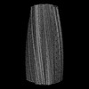
 Map data
 Sample
 Keywords
 Function and homology information
 Homo sapiens (human)
 Authors
 United Kingdom, 2 items
  Citation
 Structure visualization
 Downloads & links
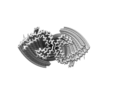 emd_19854.png
 http://ftp.pdbj.org/pub/emdb/structures/EMD-19854


 Z (Sec.)
 Y (Row.)
 X (Col.)


 Sample components
 Processing
 Electron microscopy
 FIELD EMISSION GUN