 Movie
Movie Controller

[English] 日本語
 Yorodumi
- PDB-9eea: Cryo-EM structure of the adenosine A2A receptor intermediate boun... -
+ Open data

- Basic information
| Entry | Database: PDB / ID: 9eea | |||||||||||||||||||||||||||
|---|---|---|---|---|---|---|---|---|---|---|---|---|---|---|---|---|---|---|---|---|---|---|---|---|---|---|---|---|
| Title | Cryo-EM structure of the adenosine A2A receptor intermediate bound to a miniGs heterotrimer | |||||||||||||||||||||||||||
 Components Components |
| |||||||||||||||||||||||||||
 Keywords Keywords | SIGNALING PROTEIN/IMMUNE SYSTEM / GPCR / Heterotrimer / Receptor / cryo-EM / SIGNALING PROTEIN-IMMUNE SYSTEM complex | |||||||||||||||||||||||||||
| Function / homology |  Function and homology information Function and homology informationregulation of norepinephrine secretion / positive regulation of circadian sleep/wake cycle, sleep / Adenosine P1 receptors / positive regulation of acetylcholine secretion, neurotransmission / G protein-coupled adenosine receptor activity / response to purine-containing compound / G protein-coupled adenosine receptor signaling pathway / NGF-independant TRKA activation / Surfactant metabolism / type 5 metabotropic glutamate receptor binding ...regulation of norepinephrine secretion / positive regulation of circadian sleep/wake cycle, sleep / Adenosine P1 receptors / positive regulation of acetylcholine secretion, neurotransmission / G protein-coupled adenosine receptor activity / response to purine-containing compound / G protein-coupled adenosine receptor signaling pathway / NGF-independant TRKA activation / Surfactant metabolism / type 5 metabotropic glutamate receptor binding / negative regulation of vascular permeability / positive regulation of urine volume / intermediate filament / response to caffeine / blood circulation / presynaptic active zone / synaptic transmission, cholinergic / sensory perception / eating behavior / positive regulation of glutamate secretion / regulation of calcium ion transport / adenylate cyclase-activating G protein-coupled bile acid receptor signaling pathway / adenylate cyclase-activating serotonin receptor signaling pathway / regulation of skeletal muscle contraction / alpha-actinin binding / synaptic transmission, dopaminergic / hair follicle placode formation / PKA activation in glucagon signalling / developmental growth / membrane depolarization / asymmetric synapse / intracellular transport / axolemma / D1 dopamine receptor binding / prepulse inhibition / cellular defense response / renal water homeostasis / vascular endothelial cell response to laminar fluid shear stress / phagocytosis / Hedgehog 'off' state / activation of adenylate cyclase activity / adenylate cyclase-activating adrenergic receptor signaling pathway / cellular response to acidic pH / neuron projection morphogenesis / positive regulation of synaptic transmission, glutamatergic / presynaptic modulation of chemical synaptic transmission / cellular response to glucagon stimulus / astrocyte activation / regulation of mitochondrial membrane potential / intracellular glucose homeostasis / excitatory postsynaptic potential / positive regulation of protein secretion / adenylate cyclase activator activity / trans-Golgi network membrane / positive regulation of insulin secretion involved in cellular response to glucose stimulus / locomotory behavior / positive regulation of long-term synaptic potentiation / central nervous system development / negative regulation of inflammatory response to antigenic stimulus / synaptic transmission, glutamatergic / response to prostaglandin E / bone development / vasodilation / platelet aggregation / cognition / positive regulation of insulin secretion / G-protein beta/gamma-subunit complex binding / blood coagulation / adenylate cyclase-modulating G protein-coupled receptor signaling pathway / phospholipase C-activating G protein-coupled receptor signaling pathway / Olfactory Signaling Pathway / Activation of the phototransduction cascade / G protein-coupled acetylcholine receptor signaling pathway / sensory perception of smell / G beta:gamma signalling through PLC beta / Presynaptic function of Kainate receptors / Thromboxane signalling through TP receptor / Activation of G protein gated Potassium channels / Inhibition of voltage gated Ca2+ channels via Gbeta/gamma subunits / G-protein activation / Glucagon signaling in metabolic regulation / G beta:gamma signalling through CDC42 / Prostacyclin signalling through prostacyclin receptor / Synthesis, secretion, and inactivation of Glucagon-like Peptide-1 (GLP-1) / G beta:gamma signalling through BTK / photoreceptor disc membrane / ADP signalling through P2Y purinoceptor 12 / Glucagon-type ligand receptors / Sensory perception of sweet, bitter, and umami (glutamate) taste / Adrenaline,noradrenaline inhibits insulin secretion / cell-cell signaling / Vasopressin regulates renal water homeostasis via Aquaporins / Glucagon-like Peptide-1 (GLP1) regulates insulin secretion / G alpha (z) signalling events / cellular response to catecholamine stimulus / ADP signalling through P2Y purinoceptor 1 / G beta:gamma signalling through PI3Kgamma / ADORA2B mediated anti-inflammatory cytokines production / adenylate cyclase-activating dopamine receptor signaling pathway / cellular response to prostaglandin E stimulus Similarity search - Function | |||||||||||||||||||||||||||
| Biological species |  Homo sapiens (human) Homo sapiens (human) | |||||||||||||||||||||||||||
| Method | ELECTRON MICROSCOPY / single particle reconstruction / cryo EM / Resolution: 3.36 Å | |||||||||||||||||||||||||||
 Authors Authors | Bi, M. / Wang, X. / Ye, L. / Cheng, Y. | |||||||||||||||||||||||||||
| Funding support |  United States, 2items United States, 2items
| |||||||||||||||||||||||||||
 Citation Citation | Journal: Nat Commun / Year: 2025 Title: Structure and function of a near fully-activated intermediate GPCR-Gαβγ complex. Authors: Maxine Bi / Xudong Wang / Jinan Wang / Jun Xu / Wenkai Sun / Victor Ayo Adediwura / Yinglong Miao / Yifan Cheng / Libin Ye / Abstract: Unraveling the signaling roles of intermediate complexes is pivotal for G protein-coupled receptor (GPCR) drug development. Despite hundreds of GPCR-Gαβγ structures, these snapshots primarily ...Unraveling the signaling roles of intermediate complexes is pivotal for G protein-coupled receptor (GPCR) drug development. Despite hundreds of GPCR-Gαβγ structures, these snapshots primarily capture the fully activated complex. Consequently, the functions of intermediate GPCR-G protein complexes remain elusive. Guided by a conformational landscape visualized via F quantitative NMR and molecular dynamics (MD) simulations, we determined the structure of an intermediate GPCR-mini-Gαβγ complex at 2.6 Å using cryo-EM, by blocking its transition to the fully activated complex. Furthermore, we present direct evidence that the complex at this intermediate state initiates a rate-limited nucleotide exchange before transitioning to the fully activated complex. In this state, BODIPY-GDP/GTP based nucleotide exchange assays further indicated the α-helical domain of the Gα is partially open, allowing it to grasp a nucleotide at a non-canonical binding site, distinct from the canonical nucleotide-binding site. These advances bridge a significant gap in our understanding of the complexity of GPCR signaling. | |||||||||||||||||||||||||||
| History |
|
- Structure visualization
| Structure viewer | Molecule: MolmilJmol/JSmol |
|---|
- Downloads & links
-Download
| PDBx/mmCIF format | 9eea.cif.gz | 365.9 KB | Display | PDBx/mmCIF format |
|---|---|---|---|---|
| PDB format | pdb9eea.ent.gz | 293 KB | Display | PDB format |
| PDBx/mmJSON format | 9eea.json.gz | Tree view | PDBx/mmJSON format | |
| Others |  Other downloads Other downloads |
-Validation report
| Arichive directory | https://data.pdbj.org/pub/pdb/validation_reports/ee/9eeaftp://data.pdbj.org/pub/pdb/validation_reports/ee/9eea | HTTPS FTP |
|---|
-Related structure data
| Related structure data |  47953MC  9ee8C 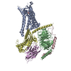 9ee9C M: map data used to model this data C: citing same article ( |
|---|---|
| Similar structure data |
-Links
 PDBj
PDBj


- Assembly
| Deposited unit | 
|
|---|---|
| 1 |
|
-Components
-Guanine nucleotide-binding protein ... , 3 types, 3 molecules BCD
| #2: Protein | Mass: 37728.152 Da / Num. of mol.: 1 Source method: isolated from a genetically manipulated source Source: (gene. exp.) Homo sapiens (human) / Gene: GNB1 / Production host:   Spodoptera frugiperda (fall armyworm) / References: UniProt: P62873 Spodoptera frugiperda (fall armyworm) / References: UniProt: P62873 |
|---|---|
| #3: Protein | Mass: 7861.143 Da / Num. of mol.: 1 Source method: isolated from a genetically manipulated source Source: (gene. exp.) Homo sapiens (human) / Gene: GNG2 / Production host: Spodoptera frugiperda (fall armyworm) / References: UniProt: P59768 |
| #4: Protein | Mass: 30776.713 Da / Num. of mol.: 1 Fragment: UNP residues 5-64,204-394,UNP residues 5-64,204-394 Source method: isolated from a genetically manipulated source Source: (gene. exp.) Homo sapiens (human) / Gene: GNAS, GNAS1, GSP / Production host:  References: UniProt: P63092, Hydrolases; Acting on acid anhydrides; Acting on GTP to facilitate cellular and subcellular movement |
-Protein / Antibody / Non-polymers , 3 types, 3 molecules AE
| #1: Protein | Mass: 39517.078 Da / Num. of mol.: 1 / Mutation: V229C, R291A Source method: isolated from a genetically manipulated source Source: (gene. exp.) Homo sapiens (human) / Gene: ADORA2A, ADORA2 / Production host:  Komagataella pastoris (fungus) / Strain (production host): SMD1163 / References: UniProt: P29274 Komagataella pastoris (fungus) / Strain (production host): SMD1163 / References: UniProt: P29274 |
|---|---|
| #5: Antibody | Mass: 16926.076 Da / Num. of mol.: 1 Source method: isolated from a genetically manipulated source Source: (gene. exp.) |
| #6: Chemical | ChemComp-ADN /  Mass: 267.241 Da / Num. of mol.: 1 / Source method: obtained synthetically / Formula: C10H13N5O4 / Feature type: SUBJECT OF INVESTIGATION Mass: 267.241 Da / Num. of mol.: 1 / Source method: obtained synthetically / Formula: C10H13N5O4 / Feature type: SUBJECT OF INVESTIGATION |
-Details
| Has ligand of interest | Y |
|---|---|
| Has protein modification | Y |
-Experimental details
-Experiment
| Experiment | Method: ELECTRON MICROSCOPY |
|---|---|
| EM experiment | Aggregation state: CELL / 3D reconstruction method: single particle reconstruction |
- Sample preparation
| Component | Name: Cryo-EM structure of the GPCR bound to a miniGs heterotrimer Type: COMPLEX / Entity ID: #1-#5 / Source: MULTIPLE SOURCES |
|---|---|
| Molecular weight | Value: 0.144 MDa / Experimental value: YES |
| Source (natural) | Organism: Homo sapiens (human) |
| Source (recombinant) | Organism: Komagataella pastoris (fungus) |
| Buffer solution | pH: 7.4 |
| Specimen | Embedding applied: NO / Shadowing applied: NO / Staining applied: NO / Vitrification applied: YES |
| Vitrification | Instrument: FEI VITROBOT MARK IV / Cryogen name: ETHANE / Humidity: 100 % / Chamber temperature: 277 K |
- Electron microscopy imaging
| Experimental equipment |  Model: Titan Krios / Image courtesy: FEI Company |
|---|---|
| Microscopy | Model: TFS KRIOS |
| Electron gun | Electron source:  FIELD EMISSION GUN / Accelerating voltage: 300 kV / Illumination mode: FLOOD BEAM FIELD EMISSION GUN / Accelerating voltage: 300 kV / Illumination mode: FLOOD BEAM |
| Electron lens | Mode: BRIGHT FIELD / Nominal defocus max: 1800 nm / Nominal defocus min: 800 nm |
| Image recording | Electron dose: 47.7 e/Å2 / Film or detector model: GATAN K3 (6k x 4k) |
- Processing
| EM software | Name: PHENIX / Category: model refinement | ||||||||||||||||||||||||
|---|---|---|---|---|---|---|---|---|---|---|---|---|---|---|---|---|---|---|---|---|---|---|---|---|---|
| CTF correction | Type: PHASE FLIPPING AND AMPLITUDE CORRECTION | ||||||||||||||||||||||||
| 3D reconstruction | Resolution: 3.36 Å / Resolution method: FSC 0.143 CUT-OFF / Num. of particles: 46863 / Symmetry type: POINT | ||||||||||||||||||||||||
| Refine LS restraints |
|
