 Movie
Movie Controller


+ Open data

- Basic information
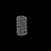
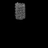
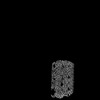
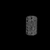
| Entry | Database: PDB / ID: 9.0E+78 | ||||||||||||
|---|---|---|---|---|---|---|---|---|---|---|---|---|---|
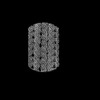
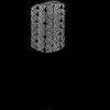
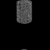
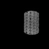
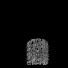
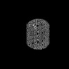
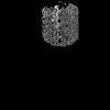
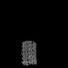
| Title | 48-nm repeat of the Leishmania tarentolae doublet microtubule | ||||||||||||
 Components Components |
| ||||||||||||
 Keywords Keywords | STRUCTURAL PROTEIN / flagella / parasite / axoneme / doublet microtubule | ||||||||||||
| Function / homology |  Function and homology information Function and homology informationouter dynein arm assembly / cilium-dependent cell motility / cilium movement / axonemal microtubule / ciliary tip / ciliary rootlet / ciliary plasm / motile cilium / cyclosporin A binding / mitotic cytokinesis ...outer dynein arm assembly / cilium-dependent cell motility / cilium movement / axonemal microtubule / ciliary tip / ciliary rootlet / ciliary plasm / motile cilium / cyclosporin A binding / mitotic cytokinesis / axoneme / cilium assembly / alpha-tubulin binding / microtubule-based process / cytoplasmic microtubule / Hsp70 protein binding / mitotic spindle organization / meiotic cell cycle / peptidylprolyl isomerase / peptidyl-prolyl cis-trans isomerase activity / Hsp90 protein binding / structural constituent of cytoskeleton / mitotic spindle / kinase activity / protein folding / microtubule / cytoskeleton / calmodulin binding / hydrolase activity / cilium / ciliary basal body / ribosome / GTPase activity / calcium ion binding / centrosome / GTP binding / metal ion binding / nucleus / membrane / cytoplasm Similarity search - Function | ||||||||||||
| Biological species |  Leishmania tarentolae (eukaryote) Leishmania tarentolae (eukaryote) | ||||||||||||
| Method | ELECTRON MICROSCOPY / single particle reconstruction / cryo EM / Resolution: 2.9 Å | ||||||||||||
 Authors Authors | Doran, M.H. / Ren, P. / Hoog, J.L. / Brown, A. | ||||||||||||
| Funding support |  United States, 3items United States, 3items
| ||||||||||||
 Citation Citation | Journal: Science / Year: 2025 Title: Evolutionary adaptations of doublet microtubules in trypanosomatid parasites. Authors: Matthew H Doran / Qingwei Niu / Jianwei Zeng / Tom Beneke / James Smith / Peter Ren / Sophia Fochler / Adrian Coscia / Johanna L Höög / Shimi Meleppattu / Polina V Lishko / Richard J ...Authors: Matthew H Doran / Qingwei Niu / Jianwei Zeng / Tom Beneke / James Smith / Peter Ren / Sophia Fochler / Adrian Coscia / Johanna L Höög / Shimi Meleppattu / Polina V Lishko / Richard J Wheeler / Eva Gluenz / Rui Zhang / Alan Brown /    Abstract: The movement and pathogenicity of trypanosomatid species, the causative agents of trypanosomiasis and leishmaniasis, are dependent on a flagellum that contains an axoneme of dynein-bound doublet ...The movement and pathogenicity of trypanosomatid species, the causative agents of trypanosomiasis and leishmaniasis, are dependent on a flagellum that contains an axoneme of dynein-bound doublet microtubules (DMTs). In this work, we present cryo-electron microscopy structures of DMTs from two trypanosomatid species, and , at resolutions up to 2.7 angstrom. The structures revealed 27 trypanosomatid-specific microtubule inner proteins, a specialized dynein-docking complex, and the presence of paralogous proteins that enable higher-order periodicities or proximal-distal patterning. Leveraging the genetic tractability of trypanosomatid species, we quantified the location and contribution of each structure-identified protein to swimming behavior. Our study shows that proper B-tubule closure is critical for flagellar motility, exemplifying how integrating structural identification with systematic gene deletion can dissect individual protein contributions to flagellar motility. | ||||||||||||
| History |
|
- Structure visualization
| Structure viewer | Molecule: MolmilJmol/JSmol |
|---|
- Downloads & links
-Download
| PDBx/mmCIF format | 9e78.cif.gz | 28.8 MB | Display | PDBx/mmCIF format |
|---|---|---|---|---|
| PDB format | pdb9e78.ent.gz | Display | PDB format | |
| PDBx/mmJSON format | 9e78.json.gz | Tree view | PDBx/mmJSON format | |
| Others |  Other downloads Other downloads |
-Validation report
| Summary document | 9e78_validation.pdf.gz | 20.7 MB | Display | wwPDB validaton report |
|---|---|---|---|---|
| Full document | 9e78_full_validation.pdf.gz | 20.8 MB | Display | |
| Data in XML | 9e78_validation.xml.gz | 2.2 MB | Display | |
| Data in CIF | 9e78_validation.cif.gz | 4.4 MB | Display | |
| Arichive directory | https://data.pdbj.org/pub/pdb/validation_reports/e7/9e78ftp://data.pdbj.org/pub/pdb/validation_reports/e7/9e78 | HTTPS FTP |
-Related structure data
| Related structure data |  47661MC C: citing same article ( M: map data used to model this data |
|---|---|
| Similar structure data |


-Links
 PDBj
PDBj


- Assembly
| Deposited unit | 
|
|---|---|
| 1 |
|
-Components
+Protein , 68 types, 491 molecules AABADAFAHAJALANBBBBDBFBHBJBLBNCACCCECGCICKCMDADCDEDGDIDKDM...
-Non-polymers , 6 types, 511 molecules 


| #69: Chemical | ChemComp-GDP /  Type: RNA linking / Mass: 443.201 Da / Num. of mol.: 161 / Source method: obtained synthetically / Formula: C10H15N5O11P2 / Comment: GDP, energy-carrying molecule*YM Type: RNA linking / Mass: 443.201 Da / Num. of mol.: 161 / Source method: obtained synthetically / Formula: C10H15N5O11P2 / Comment: GDP, energy-carrying molecule*YM#70: Chemical | ChemComp-MG /  Mass: 24.305 Da / Num. of mol.: 158 / Source method: obtained synthetically / Formula: Mg Mass: 24.305 Da / Num. of mol.: 158 / Source method: obtained synthetically / Formula: Mg#71: Chemical | ChemComp-GTP /  Mass: 523.180 Da / Num. of mol.: 158 / Source method: obtained synthetically / Formula: C10H16N5O14P3 / Comment: GTP, energy-carrying molecule*YM Mass: 523.180 Da / Num. of mol.: 158 / Source method: obtained synthetically / Formula: C10H16N5O14P3 / Comment: GTP, energy-carrying molecule*YM#72: Chemical | ChemComp-ZN /  Mass: 65.409 Da / Num. of mol.: 29 / Source method: obtained synthetically / Formula: Zn Mass: 65.409 Da / Num. of mol.: 29 / Source method: obtained synthetically / Formula: Zn#73: Chemical |  Mass: 40.078 Da / Num. of mol.: 3 / Source method: obtained synthetically / Formula: Ca Mass: 40.078 Da / Num. of mol.: 3 / Source method: obtained synthetically / Formula: Ca#74: Chemical |  Mass: 507.181 Da / Num. of mol.: 2 / Source method: obtained synthetically / Formula: C10H16N5O13P3 / Comment: ATP, energy-carrying molecule*YM Mass: 507.181 Da / Num. of mol.: 2 / Source method: obtained synthetically / Formula: C10H16N5O13P3 / Comment: ATP, energy-carrying molecule*YM |
|---|
-Details
| Has ligand of interest | N |
|---|---|
| Has protein modification | Y |
-Experimental details
-Experiment
| Experiment | Method: ELECTRON MICROSCOPY |
|---|---|
| EM experiment | Aggregation state: FILAMENT / 3D reconstruction method: single particle reconstruction |
- Sample preparation
| Component | Name: 48-nm structure of the Leishmania tarentolae doublet microtubule Type: COMPLEX / Entity ID: #1-#49, #52-#68 / Source: NATURAL |
|---|---|
| Molecular weight | Experimental value: NO |
| Source (natural) | Organism: Leishmania tarentolae (eukaryote) / Strain: P10 / Organelle: Flagella |
| Buffer solution | pH: 7.2 |
| Specimen | Conc.: 6.5 mg/ml / Embedding applied: NO / Shadowing applied: NO / Staining applied: NO / Vitrification applied: YES Details: This sample consisted of freshly splayed Leishmania tarentolae axonemes. |
| Specimen support | Grid material: COPPER / Grid mesh size: 300 divisions/in. / Grid type: Quantifoil R2/1 |
| Vitrification | Instrument: FEI VITROBOT MARK IV / Cryogen name: ETHANE / Humidity: 100 % / Chamber temperature: 278 K |
- Electron microscopy imaging
| Experimental equipment |  Model: Titan Krios / Image courtesy: FEI Company |
|---|---|
| Microscopy | Model: TFS KRIOS |
| Electron gun | Electron source:  FIELD EMISSION GUN / Accelerating voltage: 300 kV / Illumination mode: FLOOD BEAM FIELD EMISSION GUN / Accelerating voltage: 300 kV / Illumination mode: FLOOD BEAM |
| Electron lens | Mode: BRIGHT FIELD / Nominal defocus max: 2000 nm / Nominal defocus min: 500 nm / Cs: 2.7 mm |
| Specimen holder | Cryogen: NITROGEN |
| Image recording | Electron dose: 62.6 e/Å2 / Film or detector model: GATAN K3 (6k x 4k) / Num. of grids imaged: 4 / Num. of real images: 37665 |
- Processing
| EM software |
| |||||||||||||||||||||||||||
|---|---|---|---|---|---|---|---|---|---|---|---|---|---|---|---|---|---|---|---|---|---|---|---|---|---|---|---|---|
| CTF correction | Type: PHASE FLIPPING AND AMPLITUDE CORRECTION | |||||||||||||||||||||||||||
| 3D reconstruction | Resolution: 2.9 Å / Resolution method: FSC 0.143 CUT-OFF / Num. of particles: 533420 / Symmetry type: POINT | |||||||||||||||||||||||||||
| Atomic model building | Details: The initial model consisted of rigid body fit AlphaFold models and models build with ModelAngelo. Source name: Other / Type: integrative model |
