 Movie
Movie Controller

[English] 日本語
 Yorodumi
- EMDB-47641: Leishmania tarentolae 48 nm doublet microtubule protofilament 8.2... -
+ Open data

- Basic information
| Entry | 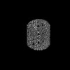 | |||||||||
|---|---|---|---|---|---|---|---|---|---|---|
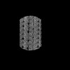
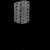
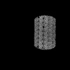
| Title | Leishmania tarentolae 48 nm doublet microtubule protofilament 8.2 focused refinement | |||||||||
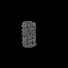
 Map data Map data | Deepemhancer map of the Leishmania tarentolae 48 nm doublet microtubule protofilament 8.2 focused refinement | |||||||||
 Sample Sample |
| |||||||||
 Keywords Keywords | flagella / parasite / axoneme / doublet microtubule / STRUCTURAL PROTEIN | |||||||||
| Biological species |  Leishmania tarentolae (eukaryote) Leishmania tarentolae (eukaryote) | |||||||||
| Method | single particle reconstruction / cryo EM / Resolution: 3.3 Å | |||||||||
 Authors Authors | Doran MH / Brown A | |||||||||
| Funding support |  United States, 2 items United States, 2 items
| |||||||||
 Citation Citation | Journal: Science / Year: 2025 Title: Evolutionary adaptations of doublet microtubules in trypanosomatid parasites. Authors: Matthew H Doran / Qingwei Niu / Jianwei Zeng / Tom Beneke / James Smith / Peter Ren / Sophia Fochler / Adrian Coscia / Johanna L Höög / Shimi Meleppattu / Polina V Lishko / Richard J ...Authors: Matthew H Doran / Qingwei Niu / Jianwei Zeng / Tom Beneke / James Smith / Peter Ren / Sophia Fochler / Adrian Coscia / Johanna L Höög / Shimi Meleppattu / Polina V Lishko / Richard J Wheeler / Eva Gluenz / Rui Zhang / Alan Brown /    Abstract: The movement and pathogenicity of trypanosomatid species, the causative agents of trypanosomiasis and leishmaniasis, are dependent on a flagellum that contains an axoneme of dynein-bound doublet ...The movement and pathogenicity of trypanosomatid species, the causative agents of trypanosomiasis and leishmaniasis, are dependent on a flagellum that contains an axoneme of dynein-bound doublet microtubules (DMTs). In this work, we present cryo-electron microscopy structures of DMTs from two trypanosomatid species, and , at resolutions up to 2.7 angstrom. The structures revealed 27 trypanosomatid-specific microtubule inner proteins, a specialized dynein-docking complex, and the presence of paralogous proteins that enable higher-order periodicities or proximal-distal patterning. Leveraging the genetic tractability of trypanosomatid species, we quantified the location and contribution of each structure-identified protein to swimming behavior. Our study shows that proper B-tubule closure is critical for flagellar motility, exemplifying how integrating structural identification with systematic gene deletion can dissect individual protein contributions to flagellar motility. | |||||||||
| History |
|
- Structure visualization
| Supplemental images |
|---|

- Downloads & links
-EMDB archive
| Map data | emd_47641.map.gz | 19.4 MB |  EMDB map data format EMDB map data format | |
|---|---|---|---|---|
| Header (meta data) | emd-47641-v30.xmlemd-47641.xml | 31.1 KB 31.1 KB | Display Display | EMDB header |
| Images |  emd_47641.png emd_47641.png | 52.9 KB | ||
| Filedesc metadata | emd-47641.cif.gz | 4.8 KB | ||
| Others | emd_47641_additional_1.map.gzemd_47641_additional_2.map.gzemd_47641_half_map_1.map.gzemd_47641_half_map_2.map.gz | 31.8 MB 470.9 MB 410.8 MB 410.8 MB | ||
| Archive directory |  http://ftp.pdbj.org/pub/emdb/structures/EMD-47641ftp://ftp.pdbj.org/pub/emdb/structures/EMD-47641 http://ftp.pdbj.org/pub/emdb/structures/EMD-47641ftp://ftp.pdbj.org/pub/emdb/structures/EMD-47641 | HTTPS FTP |
-Related structure data


-Links
| EMDB pages | EMDB (EBI/PDBe) / EMDataResource |
|---|
-Map
| File | Download / File: emd_47641.map.gz / Format: CCP4 / Size: 512 MB / Type: IMAGE STORED AS FLOATING POINT NUMBER (4 BYTES) | ||||||||||||||||||||||||||||||||||||
|---|---|---|---|---|---|---|---|---|---|---|---|---|---|---|---|---|---|---|---|---|---|---|---|---|---|---|---|---|---|---|---|---|---|---|---|---|---|
| Annotation | Deepemhancer map of the Leishmania tarentolae 48 nm doublet microtubule protofilament 8.2 focused refinement | ||||||||||||||||||||||||||||||||||||
| Projections & slices | Image control
Images are generated by Spider. | ||||||||||||||||||||||||||||||||||||
| Voxel size | X=Y=Z: 1.33 Å | ||||||||||||||||||||||||||||||||||||
| Density |
| ||||||||||||||||||||||||||||||||||||
| Symmetry | Space group: 1 | ||||||||||||||||||||||||||||||||||||
| Details | EMDB XML:
|
 Z (Sec.)
 Y (Row.)
 X (Col.)


-Supplemental data
-Additional map: Relion postprocessed map of the Leishmania tarentolae 48...
| File | emd_47641_additional_1.map | ||||||||||||
|---|---|---|---|---|---|---|---|---|---|---|---|---|---|
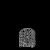
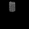
| Annotation | Relion postprocessed map of the Leishmania tarentolae 48 nm doublet microtubule protofilament 8.2 focused refinement | ||||||||||||
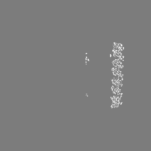
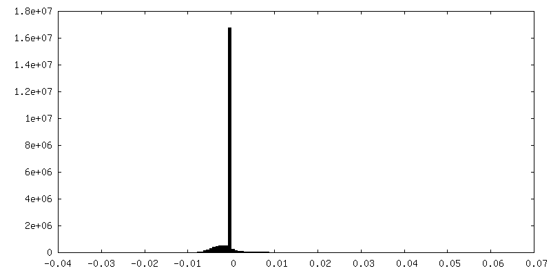
| Projections & Slices |
| ||||||||||||
| Density Histograms |


-Additional map: Relion reconstruction of the Leishmania tarentolae 48 nm...
| File | emd_47641_additional_2.map | ||||||||||||
|---|---|---|---|---|---|---|---|---|---|---|---|---|---|
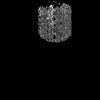
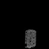
| Annotation | Relion reconstruction of the Leishmania tarentolae 48 nm doublet microtubule protofilament 8.2 focused refinement | ||||||||||||
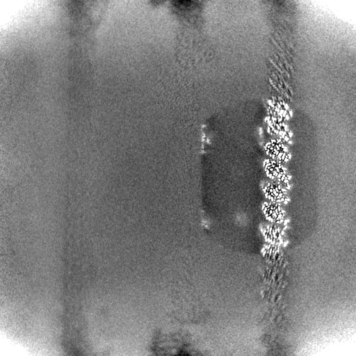
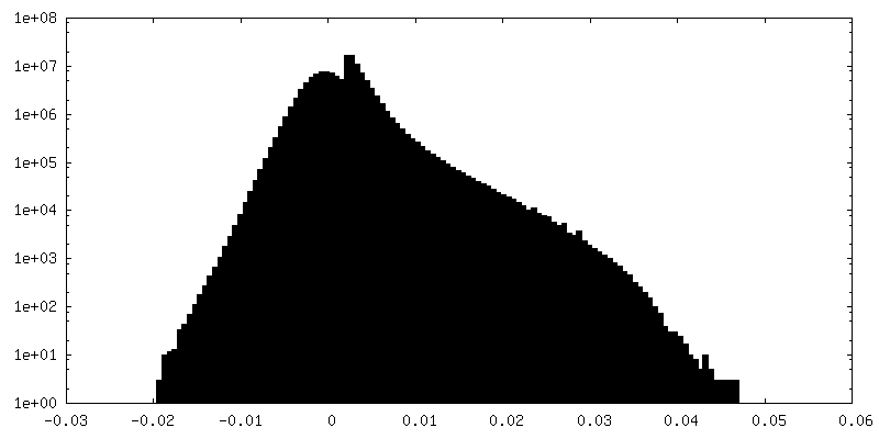
| Projections & Slices |
| ||||||||||||
| Density Histograms |


-Half map: Half map 1 of the Leishmania tarentolae 48...
| File | emd_47641_half_map_1.map | ||||||||||||
|---|---|---|---|---|---|---|---|---|---|---|---|---|---|
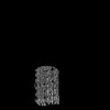
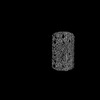
| Annotation | Half map 1 of the Leishmania tarentolae 48 nm doublet microtubule protofilament 8.2 focused refinement | ||||||||||||
| Projections & Slices |
| ||||||||||||
| Density Histograms |


-Half map: Half map 2 of the Leishmania tarentolae 48...
| File | emd_47641_half_map_2.map | ||||||||||||
|---|---|---|---|---|---|---|---|---|---|---|---|---|---|
| Annotation | Half map 2 of the Leishmania tarentolae 48 nm doublet microtubule protofilament 8.2 focused refinement | ||||||||||||
| Projections & Slices |
| ||||||||||||
| Density Histograms |


- Sample components
-Entire : 48-nm structure of the Leishmania tarentolae doublet microtubule
| Entire | Name: 48-nm structure of the Leishmania tarentolae doublet microtubule |
|---|---|
| Components |
|
-Supramolecule #1: 48-nm structure of the Leishmania tarentolae doublet microtubule
| Supramolecule | Name: 48-nm structure of the Leishmania tarentolae doublet microtubule type: complex / ID: 1 / Parent: 0 / Macromolecule list: #1-#86 |
|---|---|
| Source (natural) | Organism: Leishmania tarentolae (eukaryote) / Organelle: Flagella |
-Experimental details
-Structure determination
| Method | cryo EM |
|---|---|
 Processing Processing | single particle reconstruction |
| Aggregation state | filament |
-Sample preparation
| Concentration | 6.5 mg/mL |
|---|---|
| Buffer | pH: 7.2 |
| Grid | Model: Quantifoil R2/1 / Material: COPPER / Mesh: 300 / Support film - Material: CARBON / Support film - topology: HOLEY |
| Vitrification | Cryogen name: ETHANE / Chamber humidity: 100 % / Chamber temperature: 278 K / Instrument: FEI VITROBOT MARK IV |
| Details | This sample consisted of freshly splayed Leishmania tarentolae axonemes. |
- Electron microscopy
| Microscope | TFS KRIOS |
|---|---|
| Image recording | Film or detector model: GATAN K3 (6k x 4k) / Average electron dose: 62.6 e/Å2 |
| Electron beam | Acceleration voltage: 300 kV / Electron source:  FIELD EMISSION GUN FIELD EMISSION GUN |
| Electron optics | Illumination mode: FLOOD BEAM / Imaging mode: BRIGHT FIELD / Nominal defocus max: 2.0 µm / Nominal defocus min: 0.5 µm |
| Experimental equipment |  Model: Titan Krios / Image courtesy: FEI Company |
-Image processing
| Startup model | Type of model: EMDB MAP EMDB ID: Details: The map used for initial angular assignment is the C. reinhardtii DMT. |
|---|---|
| Final reconstruction | Resolution.type: BY AUTHOR / Resolution: 3.3 Å / Resolution method: FSC 0.143 CUT-OFF / Software - Name: RELION / Number images used: 533420 |
| Initial angle assignment | Type: MAXIMUM LIKELIHOOD / Software - Name: RELION |
| Final angle assignment | Type: MAXIMUM LIKELIHOOD / Software - Name: RELION |
-Atomic model buiding 1
| Initial model | Chain - Source name: Other / Chain - Initial model type: integrative model Details: The initial model consisted of rigid body fit AlphaFold models and models build with ModelAngelo. |
|---|
