Movie
Movie Controller

[English] 日本語
 Yorodumi
- PDB-9e19: Structure of RyR1 in the open state in the presence of pentoxifylline -
+ Open data

- Basic information
| Entry | Database: PDB / ID: 9.0E+19 | ||||||
|---|---|---|---|---|---|---|---|
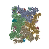
| Title | Structure of RyR1 in the open state in the presence of pentoxifylline | ||||||
 Components Components |
| ||||||
 Keywords Keywords | TRANSPORT PROTEIN / calcium channel / sarcoplasmic reticulum | ||||||
| Function / homology |  Function and homology information Function and homology informationcytoplasmic side of membrane / ATP-gated ion channel activity / terminal cisterna / ryanodine receptor complex / ryanodine-sensitive calcium-release channel activity / release of sequestered calcium ion into cytosol by sarcoplasmic reticulum / ossification involved in bone maturation / skin development / organelle membrane / cellular response to caffeine ...cytoplasmic side of membrane / ATP-gated ion channel activity / terminal cisterna / ryanodine receptor complex / ryanodine-sensitive calcium-release channel activity / release of sequestered calcium ion into cytosol by sarcoplasmic reticulum / ossification involved in bone maturation / skin development / organelle membrane / cellular response to caffeine / intracellularly gated calcium channel activity / outflow tract morphogenesis / regulation of ryanodine-sensitive calcium-release channel activity / toxic substance binding / voltage-gated calcium channel activity / smooth endoplasmic reticulum / striated muscle contraction / skeletal muscle fiber development / regulation of release of sequestered calcium ion into cytosol by sarcoplasmic reticulum / muscle contraction / sarcoplasmic reticulum membrane / release of sequestered calcium ion into cytosol / cellular response to calcium ion / sarcoplasmic reticulum / RNA polymerase II CTD heptapeptide repeat P3 isomerase activity / RNA polymerase II CTD heptapeptide repeat P6 isomerase activity / peptidylprolyl isomerase / sarcolemma / calcium ion transmembrane transport / calcium channel activity / Z disc / intracellular calcium ion homeostasis / disordered domain specific binding / protein homotetramerization / transmembrane transporter binding / calmodulin binding / intracellular membrane-bounded organelle / calcium ion binding / ATP binding / identical protein binding / membrane / cytosol Similarity search - Function | ||||||
| Biological species |  | ||||||
| Method | ELECTRON MICROSCOPY / single particle reconstruction / cryo EM / Resolution: 4.04 Å | ||||||
 Authors Authors | Miotto, M.C. / Marks, A.R. | ||||||
| Funding support | 1items
| ||||||
 Citation Citation | Journal: Proc Natl Acad Sci U S A / Year: 2025 Title: Targeting ryanodine receptors with allopurinol and xanthine derivatives for the treatment of cardiac and musculoskeletal weakness disorders. Authors: Marco C Miotto / Estefania Luna-Figueroa / Carl Tchagou / Laith Bahlouli / Steven Reiken / Haikel Dridi / Yang Liu / Gunnar Weninger / Andrew R Marks /  Abstract: Ryanodine receptors (RyRs) are intracellular Ca channels essential for muscle contraction. Caffeine, a xanthine derivative, has been known for decades to increase muscle contraction and enhance ...Ryanodine receptors (RyRs) are intracellular Ca channels essential for muscle contraction. Caffeine, a xanthine derivative, has been known for decades to increase muscle contraction and enhance activation of RyRs by increasing the sensitivity to Ca. We previously showed that xanthine, the only physiologically relevant xanthine derivative, also binds to and activates RyR2. Most xanthine derivatives and analogs are safe and widely prescribed, with the most popular being the xanthine oxidoreductase inhibitor allopurinol (~15M yearly prescriptions in USA). We propose that xanthine derivatives and analogs that enhance RyRs activity could be used for lead optimization and eventually for the treatment of the diseases that exhibit decreased muscle contraction and reduced RyRs activity, such as RyR1-related diseases, sarcopenia, and heart failure. Here, we show by cryo-EM that xanthine derivatives, analogs, and other related compounds bind to the xanthine/caffeine binding site and activate RyR1, and identify 4-oxopyrimidine as the minimal motif necessary for such interaction. | ||||||
| History |
|
- Structure visualization
| Structure viewer | Molecule: MolmilJmol/JSmol |
|---|
- Downloads & links
-Download
| PDBx/mmCIF format | 9e19.cif.gz | 3 MB | Display | PDBx/mmCIF format |
|---|---|---|---|---|
| PDB format | pdb9e19.ent.gz | Display | PDB format | |
| PDBx/mmJSON format | 9e19.json.gz | Tree view | PDBx/mmJSON format | |
| Others |  Other downloads Other downloads |
-Validation report
| Arichive directory | https://data.pdbj.org/pub/pdb/validation_reports/e1/9e19ftp://data.pdbj.org/pub/pdb/validation_reports/e1/9e19 | HTTPS FTP |
|---|
-Related structure data
| Related structure data |  47386MC  9e17C  9e18C 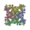 9e1aC  9e1bC  9e1cC  9e1dC  9e1eC  9e1fC  9e1gC  9e1hC  9e1iC M: map data used to model this data C: citing same article ( |
|---|---|
| Similar structure data |


-Links
 PDBj
PDBj

- Assembly
| Deposited unit | 
|
|---|---|
| 1 |
|
-Components
-Protein , 2 types, 8 molecules ABDCEHGF
| #1: Protein | Mass: 565908.625 Da / Num. of mol.: 4 / Source method: isolated from a natural source / Source: (natural) #2: Protein | Mass: 11967.705 Da / Num. of mol.: 4 / Source method: isolated from a natural source / Source: (natural) |
|---|
-Non-polymers , 4 types, 16 molecules 


| #3: Chemical | ChemComp-ATP /  Mass: 507.181 Da / Num. of mol.: 4 / Source method: obtained synthetically / Formula: C10H16N5O13P3 / Comment: ATP, energy-carrying molecule*YM Mass: 507.181 Da / Num. of mol.: 4 / Source method: obtained synthetically / Formula: C10H16N5O13P3 / Comment: ATP, energy-carrying molecule*YM#4: Chemical | ChemComp-CA /  Mass: 40.078 Da / Num. of mol.: 4 / Source method: obtained synthetically / Formula: Ca Mass: 40.078 Da / Num. of mol.: 4 / Source method: obtained synthetically / Formula: Ca#5: Chemical | ChemComp-ZN /  Mass: 65.409 Da / Num. of mol.: 4 / Source method: obtained synthetically / Formula: Zn Mass: 65.409 Da / Num. of mol.: 4 / Source method: obtained synthetically / Formula: Zn#6: Chemical | ChemComp-PNX /  Mass: 278.307 Da / Num. of mol.: 4 / Source method: obtained synthetically / Formula: C13H18N4O3 / Feature type: SUBJECT OF INVESTIGATION Mass: 278.307 Da / Num. of mol.: 4 / Source method: obtained synthetically / Formula: C13H18N4O3 / Feature type: SUBJECT OF INVESTIGATION |
|---|
-Details
| Has ligand of interest | Y |
|---|---|
| Has protein modification | Y |
-Experimental details
-Experiment
| Experiment | Method: ELECTRON MICROSCOPY |
|---|---|
| EM experiment | Aggregation state: PARTICLE / 3D reconstruction method: single particle reconstruction |
- Sample preparation
| Component |
| |||||||||||||||||||||||||||||||||||||||||||||
|---|---|---|---|---|---|---|---|---|---|---|---|---|---|---|---|---|---|---|---|---|---|---|---|---|---|---|---|---|---|---|---|---|---|---|---|---|---|---|---|---|---|---|---|---|---|---|
| Source (natural) |
| |||||||||||||||||||||||||||||||||||||||||||||
| Buffer solution | pH: 7.4 Details: 2 mM pentoxifylline was added to the final sample from a 20 mM stock solution in buffer. | |||||||||||||||||||||||||||||||||||||||||||||
| Buffer component |
| |||||||||||||||||||||||||||||||||||||||||||||
| Specimen | Conc.: 10 mg/ml / Embedding applied: NO / Shadowing applied: NO / Staining applied: NO / Vitrification applied: YES | |||||||||||||||||||||||||||||||||||||||||||||
| Vitrification | Instrument: FEI VITROBOT MARK IV / Cryogen name: ETHANE |
- Electron microscopy imaging
| Experimental equipment |  Model: Titan Krios / Image courtesy: FEI Company |
|---|---|
| Microscopy | Model: TFS KRIOS |
| Electron gun | Electron source:  FIELD EMISSION GUN / Accelerating voltage: 300 kV / Illumination mode: FLOOD BEAM FIELD EMISSION GUN / Accelerating voltage: 300 kV / Illumination mode: FLOOD BEAM |
| Electron lens | Mode: BRIGHT FIELD / Nominal defocus max: 1500 nm / Nominal defocus min: 500 nm / Cs: 2.7 mm / C2 aperture diameter: 100 µm |
| Specimen holder | Cryogen: NITROGEN / Specimen holder model: FEI TITAN KRIOS AUTOGRID HOLDER / Temperature (max): 100 K / Temperature (min): 80 K |
| Image recording | Electron dose: 58 e/Å2 / Film or detector model: GATAN K3 BIOQUANTUM (6k x 4k) |
| EM imaging optics | Energyfilter name: GIF Bioquantum / Energyfilter slit width: 20 eV |
| Image scans | Sampling size: 5 µm / Width: 5760 / Height: 4092 |
- Processing
| EM software |
| |||||||||||||||||||||||||||||||||||||||||||||
|---|---|---|---|---|---|---|---|---|---|---|---|---|---|---|---|---|---|---|---|---|---|---|---|---|---|---|---|---|---|---|---|---|---|---|---|---|---|---|---|---|---|---|---|---|---|---|
| CTF correction | Type: PHASE FLIPPING AND AMPLITUDE CORRECTION | |||||||||||||||||||||||||||||||||||||||||||||
| 3D reconstruction | Resolution: 4.04 Å / Resolution method: FSC 0.143 CUT-OFF / Num. of particles: 3336 / Symmetry type: POINT | |||||||||||||||||||||||||||||||||||||||||||||
| Atomic model building | PDB-ID: 7TZC Accession code: 7TZC / Source name: PDB / Type: experimental model | |||||||||||||||||||||||||||||||||||||||||||||
| Refine LS restraints |
|