 Movie
Movie Controller

[English] 日本語
 Yorodumi
- EMDB-47336: Structure of PKA phosphorylated human RyR2-R2474S in the open sta... -
+ Open data

- Basic information
| Entry |  | |||||||||
|---|---|---|---|---|---|---|---|---|---|---|
| Title | Structure of PKA phosphorylated human RyR2-R2474S in the open state in the presence of Calmodulin - focused map on xanthine | |||||||||
 Map data Map data | Structure of PKA phosphorylated human RyR2-R2474S in the open state in the presence of Calmodulin - focused map on xanthine | |||||||||
 Sample Sample |
| |||||||||
 Keywords Keywords | Ryanodine receptor 2 / calcium channel / MEMBRANE PROTEIN | |||||||||
| Biological species |  Homo sapiens (human) Homo sapiens (human) | |||||||||
| Method | single particle reconstruction / cryo EM / Resolution: 2.38 Å | |||||||||
 Authors Authors | Miotto MC / Marks AR | |||||||||
| Funding support |  United States, 1 items United States, 1 items
| |||||||||
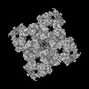
 Citation Citation | Journal: Sci Adv / Year: 2022 Title: Structural analyses of human ryanodine receptor type 2 channels reveal the mechanisms for sudden cardiac death and treatment. Authors: Marco C Miotto / Gunnar Weninger / Haikel Dridi / Qi Yuan / Yang Liu / Anetta Wronska / Zephan Melville / Leah Sittenfeld / Steven Reiken / Andrew R Marks / Abstract: Ryanodine receptor type 2 (RyR2) mutations have been linked to an inherited form of exercise-induced sudden cardiac death called catecholaminergic polymorphic ventricular tachycardia (CPVT). CPVT ...Ryanodine receptor type 2 (RyR2) mutations have been linked to an inherited form of exercise-induced sudden cardiac death called catecholaminergic polymorphic ventricular tachycardia (CPVT). CPVT results from stress-induced sarcoplasmic reticular Ca leak via the mutant RyR2 channels during diastole. We present atomic models of human wild-type (WT) RyR2 and the CPVT mutant RyR2-R2474S determined by cryo-electron microscopy with overall resolutions in the range of 2.6 to 3.6 Å, and reaching local resolutions of 2.25 Å, unprecedented for RyR2 channels. Under nonactivating conditions, the RyR2-R2474S channel is in a "primed" state between the closed and open states of WT RyR2, rendering it more sensitive to activation that results in stress-induced Ca leak. The Rycal drug ARM210 binds to RyR2-R2474S, reverting the primed state toward the closed state. Together, these studies provide a mechanism for CPVT and for the therapeutic actions of ARM210. | |||||||||
| History |
|
- Structure visualization
| Supplemental images |
|---|

- Downloads & links
-EMDB archive
| Map data | emd_47336.map.gz | 479.5 MB |  EMDB map data format EMDB map data format | |
|---|---|---|---|---|
| Header (meta data) | emd-47336-v30.xmlemd-47336.xml | 19 KB 19 KB | Display Display | EMDB header |
| Images |  emd_47336.png emd_47336.png | 24.1 KB | ||
| Filedesc metadata | emd-47336.cif.gz | 4.8 KB | ||
| Others | emd_47336_half_map_1.map.gzemd_47336_half_map_2.map.gz | 470.6 MB 470.6 MB | ||
| Archive directory |  http://ftp.pdbj.org/pub/emdb/structures/EMD-47336ftp://ftp.pdbj.org/pub/emdb/structures/EMD-47336 http://ftp.pdbj.org/pub/emdb/structures/EMD-47336ftp://ftp.pdbj.org/pub/emdb/structures/EMD-47336 | HTTPS FTP |
-Related structure data
| Related structure data |  9e17C  9e18C  9e19C 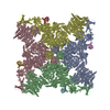 9e1aC  9e1bC  9e1cC  9e1dC  9e1eC  9e1fC  9e1gC  9e1hC  9e1iC C: citing same article ( |
|---|


-Links
| EMDB pages | EMDB (EBI/PDBe) / EMDataResource |
|---|
-Map
| File | Download / File: emd_47336.map.gz / Format: CCP4 / Size: 512 MB / Type: IMAGE STORED AS FLOATING POINT NUMBER (4 BYTES) | ||||||||||||||||||||||||||||||||||||
|---|---|---|---|---|---|---|---|---|---|---|---|---|---|---|---|---|---|---|---|---|---|---|---|---|---|---|---|---|---|---|---|---|---|---|---|---|---|
| Annotation | Structure of PKA phosphorylated human RyR2-R2474S in the open state in the presence of Calmodulin - focused map on xanthine | ||||||||||||||||||||||||||||||||||||
| Projections & slices | Image control
Images are generated by Spider. | ||||||||||||||||||||||||||||||||||||
| Voxel size | X=Y=Z: 0.83 Å | ||||||||||||||||||||||||||||||||||||
| Density |
| ||||||||||||||||||||||||||||||||||||
| Symmetry | Space group: 1 | ||||||||||||||||||||||||||||||||||||
| Details | EMDB XML:
|
 Z (Sec.)
 Y (Row.)
 X (Col.)


-Supplemental data
-Half map: #1
| File | emd_47336_half_map_1.map | ||||||||||||
|---|---|---|---|---|---|---|---|---|---|---|---|---|---|
| Projections & Slices |
| ||||||||||||
| Density Histograms |


-Half map: #2
| File | emd_47336_half_map_2.map | ||||||||||||
|---|---|---|---|---|---|---|---|---|---|---|---|---|---|
| Projections & Slices |
| ||||||||||||
| Density Histograms |


- Sample components
-Entire : Complex of RyR2-R2474S, Calstabin-2, and Calmodulin
| Entire | Name: Complex of RyR2-R2474S, Calstabin-2, and Calmodulin |
|---|---|
| Components |
|
-Supramolecule #1: Complex of RyR2-R2474S, Calstabin-2, and Calmodulin
| Supramolecule | Name: Complex of RyR2-R2474S, Calstabin-2, and Calmodulin / type: complex / ID: 1 / Parent: 0 / Macromolecule list: #3, #1-#2 |
|---|
-Supramolecule #2: Ryanodine receptor 2
| Supramolecule | Name: Ryanodine receptor 2 / type: complex / ID: 2 / Parent: 1 / Macromolecule list: #3 |
|---|---|
| Source (natural) | Organism: Homo sapiens (human) |
-Supramolecule #3: Peptidyl-prolyl cis-trans isomerase FKBP1B (E.C.5.2.1.8), Calmodulin-1
| Supramolecule | Name: Peptidyl-prolyl cis-trans isomerase FKBP1B (E.C.5.2.1.8), Calmodulin-1 type: complex / ID: 3 / Parent: 1 / Macromolecule list: #1-#2 |
|---|---|
| Source (natural) | Organism: Homo sapiens (human) |
-Experimental details
-Structure determination
| Method | cryo EM |
|---|---|
 Processing Processing | single particle reconstruction |
| Aggregation state | particle |
-Sample preparation
| Concentration | 2.2 mg/mL | |||||||||||||||||||||||||||||||||
|---|---|---|---|---|---|---|---|---|---|---|---|---|---|---|---|---|---|---|---|---|---|---|---|---|---|---|---|---|---|---|---|---|---|---|
| Buffer | pH: 7.4 Component:
Details: Xanthine was made fresh to avoid aggregation. Xanthine stock solution was 10 mM in NaOH 0.5 N. | |||||||||||||||||||||||||||||||||
| Vitrification | Cryogen name: ETHANE / Instrument: FEI VITROBOT MARK IV | |||||||||||||||||||||||||||||||||
| Details | 40 uM of Calmodulin was added to the final sample. |
- Electron microscopy
| Microscope | TFS KRIOS |
|---|---|
| Image recording | Film or detector model: GATAN K3 BIOQUANTUM (6k x 4k) / Digitization - Dimensions - Width: 5760 pixel / Digitization - Dimensions - Height: 4092 pixel / Average electron dose: 58.0 e/Å2 |
| Electron beam | Acceleration voltage: 300 kV / Electron source:  FIELD EMISSION GUN FIELD EMISSION GUN |
| Electron optics | Illumination mode: FLOOD BEAM / Imaging mode: BRIGHT FIELD / Nominal defocus max: 1.2 µm / Nominal defocus min: 0.4 µm |
| Experimental equipment |  Model: Titan Krios / Image courtesy: FEI Company |
+Image processing
-Atomic model buiding 1
| Initial model | PDB ID: Chain - Source name: PDB / Chain - Initial model type: experimental model |
|---|

