National Institutes of Health/National Institute of General Medical Sciences (NIH/NIGMS)
United States
Citation
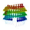
Journal: Nat Commun / Year: 2025 Title: Donor strand complementation and calcium ion coordination drive the chaperone-free polymerization of archaeal cannulae. Authors: Mike Sleutel / Ravi R Sonani / Jessalyn G Miller / Fengbin Wang / Andres Gonzalez Socorro / Yang Chen / Reece Martin / Borries Demeler / Michael J Rudolph / Vikram Alva / Han Remaut / Edward ...Authors: Mike Sleutel / Ravi R Sonani / Jessalyn G Miller / Fengbin Wang / Andres Gonzalez Socorro / Yang Chen / Reece Martin / Borries Demeler / Michael J Rudolph / Vikram Alva / Han Remaut / Edward H Egelman / Vincent P Conticello / Abstract: Cannulae are structurally rigid tubular protein filaments that accumulate on the extracellular surface of archaea within the family Pyrodictiaceae during cell growth. These obligate anaerobes ...Cannulae are structurally rigid tubular protein filaments that accumulate on the extracellular surface of archaea within the family Pyrodictiaceae during cell growth. These obligate anaerobes propagate under hyperthermophilic conditions in which cannulae form a biomatrix that interconnects and sustains cells. The persistence of cannulae in this environment suggests that these filaments display significant thermostability, which has attracted technological interest in their development as synthetic protein-based biomaterials. Here, we report cryoEM structural analyses of ex vivo and in vitro assembled recombinant cannulae. We demonstrate that the interactions between protomers in native and recombinant cannulae is based on donor strand complementation (DSC), a form of non-covalent polymerization previously observed for bacterial chaperone-usher pili. Unexpectedly, calcium ion coordination at the subunit interfaces reinforces the network of donor strand interactions in the cannulae. This study provides insight into the mechanism of assembly of cannulae and the structural origin of their high stability and rigidity.
Mass: 19989.492 Da / Num. of mol.: 1 Source method: isolated from a genetically manipulated source Source: (gene. exp.) Pyrodictium abyssi (archaea) / Production host: Escherichia coli (E. coli)
In the structure databanks used in Yorodumi, some data are registered as the other names, "COVID-19 virus" and "2019-nCoV". Here are the details of the virus and the list of structure data.
Jan 31, 2019. EMDB accession codes are about to change! (news from PDBe EMDB page)
EMDB accession codes are about to change! (news from PDBe EMDB page)
The allocation of 4 digits for EMDB accession codes will soon come to an end. Whilst these codes will remain in use, new EMDB accession codes will include an additional digit and will expand incrementally as the available range of codes is exhausted. The current 4-digit format prefixed with “EMD-” (i.e. EMD-XXXX) will advance to a 5-digit format (i.e. EMD-XXXXX), and so on. It is currently estimated that the 4-digit codes will be depleted around Spring 2019, at which point the 5-digit format will come into force.
The EM Navigator/Yorodumi systems omit the EMD- prefix.
Related info.:Q: What is EMD? / ID/Accession-code notation in Yorodumi/EM Navigator
Yorodumi is a browser for structure data from EMDB, PDB, SASBDB, etc.
This page is also the successor to EM Navigator detail page, and also detail information page/front-end page for Omokage search.
The word "yorodu" (or yorozu) is an old Japanese word meaning "ten thousand". "mi" (miru) is to see.
Related info.:EMDB / PDB / SASBDB / Comparison of 3 databanks / Yorodumi Search / Aug 31, 2016. New EM Navigator & Yorodumi / Yorodumi Papers / Jmol/JSmol / Function and homology information / Changes in new EM Navigator and Yorodumi
 Movie
Movie Controller


 Open data
 Basic information
 Components
 Keywords
 Pyrodictium abyssi (archaea)
 X-RAY DIFFRACTION /
  Authors
 United States, 1items
  Citation


 Structure visualization
 Molmil
 Downloads & links
 Other downloads
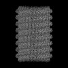


 PDBj
PDBj Assembly

 Mass: 18.015 Da / Num. of mol.: 99 / Source method: isolated from a natural source / Formula: H2O
 Sample preparation
 Processing