 Movie
Movie Controller

[English] 日本語
 Yorodumi
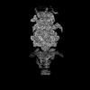
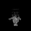
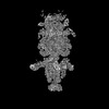
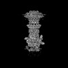
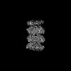
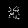
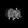
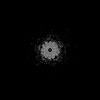
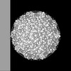
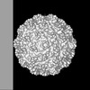
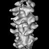
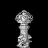
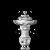
- PDB-9d9w: Mycobacteriophage Bxb1 C1 Capsid and Portal - Composite map and model -
+ Open data

- Basic information
| Entry | Database: PDB / ID: 9d9w | |||||||||||||||||||||||||||
|---|---|---|---|---|---|---|---|---|---|---|---|---|---|---|---|---|---|---|---|---|---|---|---|---|---|---|---|---|
| Title | Mycobacteriophage Bxb1 C1 Capsid and Portal - Composite map and model | |||||||||||||||||||||||||||
 Components Components |
| |||||||||||||||||||||||||||
 Keywords Keywords | VIRAL PROTEIN / Bacteriophage / Mycobacteriophage Bxb1 / capsid / portal / VIRUS | |||||||||||||||||||||||||||
| Function / homology | Portal protein / Phage portal protein, SPP1 Gp6-like / : / Phage capsid / Phage capsid family / virion component / Major capsid protein / Portal protein Function and homology information Function and homology information | |||||||||||||||||||||||||||
| Biological species |  Mycobacterium phage Bxb1 (virus) Mycobacterium phage Bxb1 (virus) | |||||||||||||||||||||||||||
| Method | ELECTRON MICROSCOPY / single particle reconstruction / cryo EM / Resolution: 3.5 Å | |||||||||||||||||||||||||||
 Authors Authors | Freeman, K.G. | |||||||||||||||||||||||||||
| Funding support |  United States, United States,  Taiwan, 8items Taiwan, 8items
| |||||||||||||||||||||||||||
 Citation Citation | Journal: Cell / Year: 2025 Title: Structure and infection dynamics of mycobacteriophage Bxb1. Authors: Krista G Freeman / Sudipta Mondal / Lourriel S Macale / Jennifer Podgorski / Simon J White / Benjamin H Silva / Valery Ortiz / Alexis Huet / Ronelito J Perez / Joemark T Narsico / Meng-Chiao ...Authors: Krista G Freeman / Sudipta Mondal / Lourriel S Macale / Jennifer Podgorski / Simon J White / Benjamin H Silva / Valery Ortiz / Alexis Huet / Ronelito J Perez / Joemark T Narsico / Meng-Chiao Ho / Deborah Jacobs-Sera / Todd L Lowary / James F Conway / Donghyun Park / Graham F Hatfull / Abstract: Mycobacteriophage Bxb1 is a well-characterized virus of Mycobacterium smegmatis with double-stranded DNA and a long, flexible tail. Mycobacteriophages show considerable potential as therapies for ...Mycobacteriophage Bxb1 is a well-characterized virus of Mycobacterium smegmatis with double-stranded DNA and a long, flexible tail. Mycobacteriophages show considerable potential as therapies for Mycobacterium infections, but little is known about the structural details of these phages or how they bind to and traverse the complex Mycobacterium cell wall. Here, we report the complete structure and atomic model of phage Bxb1, including the arrangement of immunodominant domains of both the capsid and tail tube subunits, as well as the assembly of the protein subunits in the tail-tip complex. The structure contains protein assemblies with 3-, 5-, 6-, and 12-fold symmetries, which interact to satisfy several symmetry mismatches. Cryoelectron tomography of phage particles bound to M. smegmatis reveals the structural transitions that occur for free phage particles to bind to the cell surface and navigate through the cell wall to enable DNA transfer into the cytoplasm. | |||||||||||||||||||||||||||
| History |
|
- Structure visualization
| Structure viewer | Molecule: MolmilJmol/JSmol |
|---|
- Downloads & links
-Download
| PDBx/mmCIF format | 9d9w.cif.gz | 5.5 MB | Display | PDBx/mmCIF format |
|---|---|---|---|---|
| PDB format | pdb9d9w.ent.gz | Display | PDB format | |
| PDBx/mmJSON format | 9d9w.json.gz | Tree view | PDBx/mmJSON format | |
| Others |  Other downloads Other downloads |
-Validation report
| Arichive directory | https://data.pdbj.org/pub/pdb/validation_reports/d9/9d9wftp://data.pdbj.org/pub/pdb/validation_reports/d9/9d9w | HTTPS FTP |
|---|
-Related structure data
| Related structure data | 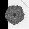 46681MC  9d93C  9d94C 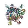 9d9lC 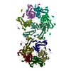 9d9xC C: citing same article ( M: map data used to model this data |
|---|---|
| Similar structure data |


-Links
 PDBj
PDBj

- Assembly
| Deposited unit | 
|
|---|---|
| 1 |
|
-Components
| #1: Protein | Mass: 41875.461 Da / Num. of mol.: 30 / Source method: isolated from a natural source / Source: (natural) Mycobacterium phage Bxb1 (virus) / References: UniProt: Q9B0A7#2: Protein | Mass: 53822.113 Da / Num. of mol.: 12 / Source method: isolated from a natural source / Source: (natural) Mycobacterium phage Bxb1 (virus) / References: UniProt: Q9B0B0Has protein modification | N | |
|---|
-Experimental details
-Experiment
| Experiment | Method: ELECTRON MICROSCOPY |
|---|---|
| EM experiment | Aggregation state: PARTICLE / 3D reconstruction method: single particle reconstruction |
- Sample preparation
| Component | Name: Mycobacterium phage Bxb1 / Type: VIRUS Details: Portal and connector complex of Bxb1, containing five protein subunit types. This is a composite map, and related entries for consensus and locally refined maps are noted. Entity ID: all / Source: NATURAL | |||||||||||||||||||||||||
|---|---|---|---|---|---|---|---|---|---|---|---|---|---|---|---|---|---|---|---|---|---|---|---|---|---|---|
| Molecular weight |
| |||||||||||||||||||||||||
| Source (natural) | Organism: Mycobacterium phage Bxb1 (virus) / Strain: Mycobacterium phage Bxb1 | |||||||||||||||||||||||||
| Details of virus | Empty: NO / Enveloped: NO / Isolate: SPECIES / Type: VIRION | |||||||||||||||||||||||||
| Natural host | Organism: Mycolicibacterium smegmatis MC2 155 | |||||||||||||||||||||||||
| Buffer solution | pH: 7.5 | |||||||||||||||||||||||||
| Buffer component |
| |||||||||||||||||||||||||
| Specimen | Conc.: 10 mg/ml / Embedding applied: NO / Shadowing applied: NO / Staining applied: NO / Vitrification applied: YES | |||||||||||||||||||||||||
| Vitrification | Instrument: FEI VITROBOT MARK IV / Cryogen name: ETHANE / Humidity: 100 % / Chamber temperature: 283.15 K |
- Electron microscopy imaging
| Experimental equipment |  Model: Titan Krios / Image courtesy: FEI Company |
|---|---|
| Microscopy | Model: FEI TITAN KRIOS |
| Electron gun | Electron source:  FIELD EMISSION GUN / Accelerating voltage: 300 kV / Illumination mode: FLOOD BEAM FIELD EMISSION GUN / Accelerating voltage: 300 kV / Illumination mode: FLOOD BEAM |
| Electron lens | Mode: BRIGHT FIELD / Nominal defocus max: 2000 nm / Nominal defocus min: 800 nm |
| Image recording | Electron dose: 30 e/Å2 / Film or detector model: GATAN K3 (6k x 4k) |
- Processing
| EM software | Name: PHENIX / Version: 1.20.1_4487: / Category: model refinement | ||||||||||||||||||||||||
|---|---|---|---|---|---|---|---|---|---|---|---|---|---|---|---|---|---|---|---|---|---|---|---|---|---|
| CTF correction | Type: PHASE FLIPPING AND AMPLITUDE CORRECTION | ||||||||||||||||||||||||
| Symmetry | Point symmetry: C1 (asymmetric) | ||||||||||||||||||||||||
| 3D reconstruction | Resolution: 3.5 Å / Resolution method: OTHER / Num. of particles: 33132 / Symmetry type: POINT | ||||||||||||||||||||||||
| Refine LS restraints |
|
