 ムービー
ムービー コントローラー


+ データを開く

- 基本情報
基本情報
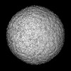
| 登録情報 | データベース: PDB / ID: 9cyt | ||||||
|---|---|---|---|---|---|---|---|
| タイトル | Cryo-EM structure of MRV outer shell | ||||||
 要素 要素 |
| ||||||
 キーワード キーワード | VIRAL PROTEIN / Mammalian reovirus / outer shell | ||||||
| 機能・相同性 |  機能・相同性情報 機能・相同性情報icosahedral viral capsid / host cell surface binding / symbiont-mediated suppression of host PKR/eIFalpha signaling / viral outer capsid / symbiont entry into host cell via permeabilization of host membrane / host cell endoplasmic reticulum / protein serine/threonine kinase inhibitor activity / host cell mitochondrion / viral life cycle / regulation of translation ...icosahedral viral capsid / host cell surface binding / symbiont-mediated suppression of host PKR/eIFalpha signaling / viral outer capsid / symbiont entry into host cell via permeabilization of host membrane / host cell endoplasmic reticulum / protein serine/threonine kinase inhibitor activity / host cell mitochondrion / viral life cycle / regulation of translation / mRNA guanylyltransferase activity / mRNA guanylyltransferase / mRNA (guanine-N7)-methyltransferase / host cell cytoplasm / mRNA 5'-cap (guanine-N7-)-methyltransferase activity / symbiont-mediated suppression of host innate immune response / symbiont-mediated suppression of host type I interferon-mediated signaling pathway / GTP binding / host cell nucleus / host cell plasma membrane / structural molecule activity / RNA binding / zinc ion binding / ATP binding / membrane 類似検索 - 分子機能 | ||||||
| 生物種 |  Mammalian orthoreovirus 3 Dearing (ウイルス) Mammalian orthoreovirus 3 Dearing (ウイルス) | ||||||
| 手法 | 電子顕微鏡法 / 単粒子再構成法 / クライオ電子顕微鏡法 / 解像度: 3.7 Å | ||||||
 データ登録者 データ登録者 | Liu, X.Y. / Xia, X. / Martynowycz, M.W. / Gonen, T. / Zhou, Z.H. | ||||||
| 資金援助 |  米国, 1件 米国, 1件
| ||||||
 引用 引用 | ジャーナル: Nat Commun / 年: 2024 タイトル: Molecular sociology of virus-induced cellular condensates supporting reovirus assembly and replication. 著者: Xiaoyu Liu / Xian Xia / Michael W Martynowycz / Tamir Gonen / Z Hong Zhou / 要旨: Virus-induced cellular condensates, or viral factories, are poorly understood high-density phases where replication of many viruses occurs. Here, by cryogenic electron tomography (cryoET) of focused ...Virus-induced cellular condensates, or viral factories, are poorly understood high-density phases where replication of many viruses occurs. Here, by cryogenic electron tomography (cryoET) of focused ion beam (FIB) milling-produced lamellae of mammalian reovirus (MRV)-infected cells, we visualized the molecular organization and interplay (i.e., "molecular sociology") of host and virus in 3D at two time points post-infection, enabling a detailed description of these condensates and a mechanistic understanding of MRV replication within them. Expanding over time, the condensate fashions host ribosomes at its periphery, and host microtubules, lipid membranes, and viral molecules in its interior, forming a 3D architecture that supports the dynamic processes of viral genome replication and capsid assembly. A total of six MRV assembly intermediates are identified inside the condensate: star core, empty and genome-containing cores, empty and full virions, and outer shell particle. Except for star core, these intermediates are visualized at atomic resolution by cryogenic electron microscopy (cryoEM) of cellular extracts. The temporal sequence and spatial rearrangement among these viral intermediates choreograph the viral life cycle within the condensates. Together, the molecular sociology of MRV-induced cellular condensate highlights the functional advantage of transient enrichment of molecules at the right location and time for viral replication. | ||||||
| 履歴 |
|
- 構造の表示
構造の表示
| 構造ビューア | 分子: MolmilJmol/JSmol |
|---|
- ダウンロードとリンク
-ダウンロード
| PDBx/mmCIF形式 | 9cyt.cif.gz | 773.5 KB | 表示 | PDBx/mmCIF形式 |
|---|---|---|---|---|
| PDB形式 | pdb9cyt.ent.gz | 614.1 KB | 表示 | PDB形式 |
| PDBx/mmJSON形式 | 9cyt.json.gz | ツリー表示 | PDBx/mmJSON形式 | |
| その他 |  その他のダウンロード その他のダウンロード |
-検証レポート
| 文書・要旨 | 9cyt_validation.pdf.gz | 1.7 MB | 表示 | wwPDB検証レポート |
|---|---|---|---|---|
| 文書・詳細版 | 9cyt_full_validation.pdf.gz | 1.8 MB | 表示 | |
| XML形式データ | 9cyt_validation.xml.gz | 116.6 KB | 表示 | |
| CIF形式データ | 9cyt_validation.cif.gz | 176.2 KB | 表示 | |
| アーカイブディレクトリ | https://data.pdbj.org/pub/pdb/validation_reports/cy/9cytftp://data.pdbj.org/pub/pdb/validation_reports/cy/9cyt | HTTPS FTP |
-関連構造データ


-リンク
 PDBj
PDBj


- 集合体
集合体
| 登録構造単位 | 
|
|---|---|
| 1 |
|
-要素
| #1: タンパク質 | 分子量: 144098.766 Da / 分子数: 1 / 由来タイプ: 天然 由来: (天然) Mammalian orthoreovirus 3 Dearing (ウイルス)細胞株: LLC-MK2 参照: UniProt: P11079, mRNA guanylyltransferase, mRNA (guanine-N7)-methyltransferase | ||||
|---|---|---|---|---|---|
| #2: タンパク質 | 分子量: 76334.273 Da / 分子数: 6 / 由来タイプ: 天然 由来: (天然) Mammalian orthoreovirus 3 Dearing (ウイルス)参照: UniProt: P11078 #3: タンパク質 | 分子量: 41168.121 Da / 分子数: 3 / 由来タイプ: 天然 由来: (天然) Mammalian orthoreovirus 3 Dearing (ウイルス)参照: UniProt: P03527 Has protein modification | N | |
-実験情報
-実験
| 実験 | 手法: 電子顕微鏡法 |
|---|---|
| EM実験 | 試料の集合状態: PARTICLE / 3次元再構成法: 単粒子再構成法 |
- 試料調製
試料調製
| 構成要素 | 名称: mammalian reovirus / タイプ: VIRUS / Entity ID: all / 由来: NATURAL |
|---|---|
| 由来(天然) | 生物種: Mammalian orthoreovirus 3 Dearing (ウイルス) |
| ウイルスについての詳細 | 中空か: YES / エンベロープを持つか: NO / 単離: STRAIN / タイプ: VIRUS-LIKE PARTICLE |
| 天然宿主 | 生物種: LLC-MK2 |
| 緩衝液 | pH: 7.4 / 詳細: Phosphate-buffered saline |
| 試料 | 包埋: NO / シャドウイング: NO / 染色: NO / 凍結: YES |
| 試料支持 | グリッドの材料: GOLD / グリッドのサイズ: 300 divisions/in. グリッドのタイプ: PELCO Ultrathin Carbon with Lacey Carbon |
| 急速凍結 | 凍結剤: ETHANE / 湿度: 100 % |
- 電子顕微鏡撮影
| 実験機器 |  モデル: Titan Krios / 画像提供: FEI Company |
|---|---|
| 顕微鏡 | モデル: FEI TITAN KRIOS |
| 電子銃 | 電子線源:  FIELD EMISSION GUN / 加速電圧: 300 kV / 照射モード: FLOOD BEAM FIELD EMISSION GUN / 加速電圧: 300 kV / 照射モード: FLOOD BEAM |
| 電子レンズ | モード: BRIGHT FIELD / 倍率(公称値): 81000 X / 最大 デフォーカス(公称値): 2600 nm / 最小 デフォーカス(公称値): 1800 nm / Cs: 2.7 mm / C2レンズ絞り径: 50 µm / アライメント法: COMA FREE |
| 試料ホルダ | 凍結剤: NITROGEN 試料ホルダーモデル: FEI TITAN KRIOS AUTOGRID HOLDER |
| 撮影 | 平均露光時間: 2 sec. / 電子線照射量: 50 e/Å2 フィルム・検出器のモデル: GATAN K3 BIOQUANTUM (6k x 4k) 撮影したグリッド数: 1 / 実像数: 12101 |
| 電子光学装置 | エネルギーフィルター名称: GIF Quantum LS / エネルギーフィルタースリット幅: 20 eV |
- 解析
解析
| EMソフトウェア |
| ||||||||||||||||||||||||
|---|---|---|---|---|---|---|---|---|---|---|---|---|---|---|---|---|---|---|---|---|---|---|---|---|---|
| CTF補正 | タイプ: PHASE FLIPPING ONLY | ||||||||||||||||||||||||
| 粒子像の選択 | 選択した粒子像数: 39141 | ||||||||||||||||||||||||
| 対称性 | 点対称性: C5 (5回回転対称) | ||||||||||||||||||||||||
| 3次元再構成 | 解像度: 3.7 Å / 解像度の算出法: FSC 0.143 CUT-OFF / 粒子像の数: 8911 / クラス平均像の数: 1 / 対称性のタイプ: POINT | ||||||||||||||||||||||||
| 原子モデル構築 | プロトコル: FLEXIBLE FIT / 空間: REAL | ||||||||||||||||||||||||
| 拘束条件 |
|
