 Movie
Movie Controller


+ Open data

- Basic information
| Entry |  | |||||||||
|---|---|---|---|---|---|---|---|---|---|---|
| Title | Cryo-EM structure of MRV full core | |||||||||
 Map data Map data | map of MRV core | |||||||||
 Sample Sample |
| |||||||||
 Keywords Keywords | Mammalian reovirus / outer shell / VIRAL PROTEIN | |||||||||
| Function / homology |  Function and homology information Function and homology informationicosahedral viral capsid / T=2 icosahedral viral capsid / viral inner capsid / viral outer capsid / 7-methylguanosine mRNA capping / mRNA guanylyltransferase / mRNA guanylyltransferase activity / mRNA (guanine-N7)-methyltransferase / mRNA 5'-cap (guanine-N7-)-methyltransferase activity / RNA helicase activity ...icosahedral viral capsid / T=2 icosahedral viral capsid / viral inner capsid / viral outer capsid / 7-methylguanosine mRNA capping / mRNA guanylyltransferase / mRNA guanylyltransferase activity / mRNA (guanine-N7)-methyltransferase / mRNA 5'-cap (guanine-N7-)-methyltransferase activity / RNA helicase activity / RNA helicase / GTP binding / ATP hydrolysis activity / zinc ion binding / ATP binding Similarity search - Function | |||||||||
| Biological species |  Mammalian orthoreovirus 3 Dearing Mammalian orthoreovirus 3 Dearing | |||||||||
| Method | single particle reconstruction / cryo EM / Resolution: 3.3 Å | |||||||||
 Authors Authors | Liu XY / Xia X / Martynowycz MW / Gonen T / Zhou ZH | |||||||||
| Funding support |  United States, 1 items United States, 1 items
| |||||||||
 Citation Citation | Journal: Nat Commun / Year: 2024 Title: Molecular sociology of virus-induced cellular condensates supporting reovirus assembly and replication. Authors: Xiaoyu Liu / Xian Xia / Michael W Martynowycz / Tamir Gonen / Z Hong Zhou / Abstract: Virus-induced cellular condensates, or viral factories, are poorly understood high-density phases where replication of many viruses occurs. Here, by cryogenic electron tomography (cryoET) of focused ...Virus-induced cellular condensates, or viral factories, are poorly understood high-density phases where replication of many viruses occurs. Here, by cryogenic electron tomography (cryoET) of focused ion beam (FIB) milling-produced lamellae of mammalian reovirus (MRV)-infected cells, we visualized the molecular organization and interplay (i.e., "molecular sociology") of host and virus in 3D at two time points post-infection, enabling a detailed description of these condensates and a mechanistic understanding of MRV replication within them. Expanding over time, the condensate fashions host ribosomes at its periphery, and host microtubules, lipid membranes, and viral molecules in its interior, forming a 3D architecture that supports the dynamic processes of viral genome replication and capsid assembly. A total of six MRV assembly intermediates are identified inside the condensate: star core, empty and genome-containing cores, empty and full virions, and outer shell particle. Except for star core, these intermediates are visualized at atomic resolution by cryogenic electron microscopy (cryoEM) of cellular extracts. The temporal sequence and spatial rearrangement among these viral intermediates choreograph the viral life cycle within the condensates. Together, the molecular sociology of MRV-induced cellular condensate highlights the functional advantage of transient enrichment of molecules at the right location and time for viral replication. | |||||||||
| History |
|
- Structure visualization
| Supplemental images |
|---|

- Downloads & links
-EMDB archive
| Map data | emd_46053.map.gz | 201.1 MB | EMDB map data format | |
|---|---|---|---|---|
| Header (meta data) | emd-46053-v30.xmlemd-46053.xml | 22.8 KB 22.8 KB | Display Display | EMDB header |
| Images |  emd_46053.png emd_46053.png | 122.3 KB | ||
| Filedesc metadata | emd-46053.cif.gz | 7.9 KB | ||
| Others | emd_46053_half_map_1.map.gzemd_46053_half_map_2.map.gz | 170.9 MB 171 MB | ||
| Archive directory |  http://ftp.pdbj.org/pub/emdb/structures/EMD-46053ftp://ftp.pdbj.org/pub/emdb/structures/EMD-46053 http://ftp.pdbj.org/pub/emdb/structures/EMD-46053ftp://ftp.pdbj.org/pub/emdb/structures/EMD-46053 | HTTPS FTP |
-Related structure data
| Related structure data |  9cyxMC 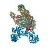 9cytC  9cyyC M: atomic model generated by this map C: citing same article ( |
|---|---|
| Similar structure data |


-Links
| EMDB pages | EMDB (EBI/PDBe) / EMDataResource |
|---|---|
| Related items in Molecule of the Month |


-Map
| File | Download / File: emd_46053.map.gz / Format: CCP4 / Size: 216 MB / Type: IMAGE STORED AS FLOATING POINT NUMBER (4 BYTES) | ||||||||||||||||||||||||||||||||||||
|---|---|---|---|---|---|---|---|---|---|---|---|---|---|---|---|---|---|---|---|---|---|---|---|---|---|---|---|---|---|---|---|---|---|---|---|---|---|
| Annotation | map of MRV core | ||||||||||||||||||||||||||||||||||||
| Projections & slices | Image control
Images are generated by Spider. | ||||||||||||||||||||||||||||||||||||
| Voxel size | X=Y=Z: 1.1 Å | ||||||||||||||||||||||||||||||||||||
| Density |
| ||||||||||||||||||||||||||||||||||||
| Symmetry | Space group: 1 | ||||||||||||||||||||||||||||||||||||
| Details | EMDB XML:
|
 Z (Sec.)
 Y (Row.)
 X (Col.)


-Supplemental data
-Half map: half2 map of MRV core
| File | emd_46053_half_map_1.map | ||||||||||||
|---|---|---|---|---|---|---|---|---|---|---|---|---|---|
| Annotation | half2 map of MRV core | ||||||||||||
| Projections & Slices |
| ||||||||||||
| Density Histograms |


-Half map: half1 map of MRV core
| File | emd_46053_half_map_2.map | ||||||||||||
|---|---|---|---|---|---|---|---|---|---|---|---|---|---|
| Annotation | half1 map of MRV core | ||||||||||||
| Projections & Slices |
| ||||||||||||
| Density Histograms |


- Sample components
-Entire : Mammalian orthoreovirus 3 Dearing
| Entire | Name: Mammalian orthoreovirus 3 Dearing |
|---|---|
| Components |
|
-Supramolecule #1: Mammalian orthoreovirus 3 Dearing
| Supramolecule | Name: Mammalian orthoreovirus 3 Dearing / type: virus / ID: 1 / Parent: 0 / Macromolecule list: all / NCBI-ID: 10886 / Sci species name: Mammalian orthoreovirus 3 Dearing / Virus type: VIRION / Virus isolate: STRAIN / Virus enveloped: No / Virus empty: No |
|---|---|
| Host (natural) | Organism: LLC-MK2 |
-Macromolecule #1: Lambda 1
| Macromolecule | Name: Lambda 1 / type: protein_or_peptide / ID: 1 / Number of copies: 3 / Enantiomer: LEVO |
|---|---|
| Source (natural) | Organism: Mammalian orthoreovirus 3 Dearing |
| Molecular weight | Theoretical: 141.937375 KDa |
| Sequence | String: MKRIPRKTKG KSSGKGNDST ERADDGSSQL RDKQNNKAGP ATTEPGTSNR EQYKARPGIA SVQRATESAE MPMKNNDEGT PDKKGNTKG DLVNEHSEAK DEADEATKKQ AKDTDKSKAQ VTYSDTGINN ANELSRSGNV DNEGGSNQKP MSTRIAEATS A IVSKHPAR ...String: MKRIPRKTKG KSSGKGNDST ERADDGSSQL RDKQNNKAGP ATTEPGTSNR EQYKARPGIA SVQRATESAE MPMKNNDEGT PDKKGNTKG DLVNEHSEAK DEADEATKKQ AKDTDKSKAQ VTYSDTGINN ANELSRSGNV DNEGGSNQKP MSTRIAEATS A IVSKHPAR VGLPPTASSG HGYQCHVCSA VLFSPLDLDA HVASHGLHGN MTLTSSDIQR HITEFISSWQ NHPIVQVSAD VE NKKTAQL LHADTPRLVT WDAGLCTSFK IVPIVPAQVP QDVLAYTFFT SSYAIQSPFP EAAVSRIVVH TRWASNVDFD RDS SVIMAP PTENNIHLFK QLLNTETLSV RGANPLMFRA NVLHMLLEFV LDNLYLNRHT GFSQDHTPFT EGANLRSLPG PDAE KWYSI MYPTRMGTPN VSKICNFVAS CVRNRVGRFD RAQMMNGAMS EWVDVFETSD ALTVSIRGRW MARLARMNIN PTEIE WALT ECAQGYVTVT SPYAPSVNRL MPYRISNAER QISQIIRIMN IGNNATVIQP VLQDISVLLQ RISPLQIDPT IISNTM STV SESTTQTLSP ASSILGKLRP SNSDFSSFRV ALAGWLYNGV VTTVIDDSSY PKDGGSVTSL ENLWDFFILA LALPLTT DP CAPVKAFMTL ANMMVGFETI PMDNQIYTQS RRASAFSTPH TWPRCFMNIQ LISPIDAPIL RQWAEIIHRY WPNPSQIR Y GAPNVFGSAN LFTPPEVLLL PIDHQPANVT TPTLDFTNEL TNWRARVCEL MKNLVDNQRY QPGWTQSLVS SMRGTLDKL KLIKSMTPMY LQQLAPVELA VIAPMLPFPP FQVPYVRLDR DRVPTMVGVT RQSRDTITQP ALSLSTTNTT VGVPLALDAR AITVALLSG KYPPDLVTNV WYADAIYPMY ADTEVFSNLQ RDMITCEAVQ TLVTLVAQIS ETQYPVDRYL DWIPSLRASA A TAATFAEW VNTSMKTAFD LSDMLLEPLL SGDPRMTQLA IQYQQYNGRT FNIIPEMPGS VIADCVQLTA EVFNHEYNLF GI ARGDIII GRVQSTHLWS PLAPPPDLVF DRDTPGVHIF GRDCRISFGM NGAAPMIRDE TGLMVPFEGN WIFPLALWQM NTR YFNQQF DAWIKTGELR IRIEMGAYPY MLHYYDPRQY ANAWNLTSAW LEEITPTSIP SVPFMVPISS DHDISSAPAV QYII STEYN DRSLFCTNSS SPQTIAGPDK HIPVERYNIL TNPDAPPTQI QLPEVVDLYN VVTRYAYETP PITAVVMGVP UniProtKB: Inner capsid protein lambda-1 |
-Macromolecule #2: Outer capsid protein lambda-2
| Macromolecule | Name: Outer capsid protein lambda-2 / type: protein_or_peptide / ID: 2 / Number of copies: 1 / Enantiomer: LEVO / EC number: mRNA guanylyltransferase |
|---|---|
| Source (natural) | Organism: Mammalian orthoreovirus 3 Dearing |
| Molecular weight | Theoretical: 143.967562 KDa |
| Sequence | String: ANVWGVRLAD SLSSPTIETR TRQYTLHDLC SDLDANPGRE PWKPLRNQRT NNIVAVQLFR PLQGLVLDTQ LYGFPGAFDD WERFMREKL RVLKYEVLRI YPISNYSNEH VNVFVANALV GAFLSNQAFY DLLPLLIIND TMIGDLLGTG ASLSQFFQSH G DVLEVAAG ...String: ANVWGVRLAD SLSSPTIETR TRQYTLHDLC SDLDANPGRE PWKPLRNQRT NNIVAVQLFR PLQGLVLDTQ LYGFPGAFDD WERFMREKL RVLKYEVLRI YPISNYSNEH VNVFVANALV GAFLSNQAFY DLLPLLIIND TMIGDLLGTG ASLSQFFQSH G DVLEVAAG RKYLQMENYS NDDDDPPLFA KDLSDYAKAF YSDTYEVLDR FFWTHDSSAG VLVHYDKPTN GHHYLLGTLT QM VSAPPYI INATDAMLLE SCLEQFSANV RARPAQPVTR LDQCYHLRWG AQYVGEDSLT YRLGVLSLLA TNGYQLARPI PRQ LTNRWL SSFVSQIMSD GVNETPLWPQ ERYVQIAYDS PSVVDGATQY GYVRKNQLRL GMRISALQSL SDTPSPVQWL PQYT IDQAA MDEGDLMVSR LTQLPLRPDY GNIWVGDALS YYVDYNRSHR VVLSSELPQL PDTYFDGDEQ YGRSLFSLAR KIGDR SLVK DTAVLKHAYQ AIDPNTGKEY LRSRQSVAYF GASAGHSGAD QPLVIEPWIQ GKISGVPPPS SVRQFGYDVA RGAIVD LAR PFPSGDYQFV YSDVDQVVDG HDDLSISSGL VESLLSSCMH ATAPGGSFVV KINFPTRPVW HYIEQKILPN ITSYMLI KP FVTNNVELFF VAFGVHQHSS LTWTSGVYFF LVDHFYRYET LSTISRQLPS FGYVDDGSSV TGIETISIEN PGFSNMTQ A ARIGISGLCA NVGNARKSIA IYESHGARVL TITSRRSPAS ARRKSRLRYL PLIDPRSLEV QARTILPADP VLFENVSGA SPHVCLTMMY NFEVSSAVYD GDVVLDLGTG PEAKILELIP ATSPVTCVDI RPTAQPSGCW NVRTTFLELD YLSDGWITGV RGDIVTCML SLGAAAAGKS MTFDAAFQQL IKVLSKSTAN VVLVQVNCPT DVVRSIKGYL EIDSTNKRYR FPKFGRDEPY S DMDALEKI CRTAWPNCSI TWVPLSYDLR WTRLALLEST TLSSASIRIA ELMYKYMPIM RIDIHGLPME KRGNFIVGQN CS LVIPGFN AQDVFNCYFN SALAFSTEDV NAAMIPQVSA QFDATKGEWT LDMVFSDAGI YTMQALVGSN ANPVSLGSFV VDS PDVDIT DAWPAQLDFT IAGTDVDITV NPYYRLMTFV RIDGQWQIAN PDKFQFFSSA SGTLVMNVKL DIADKYLLYY IRDV QSRDV GFYIQHPLQL LNTITLPTNE DLFLSAPDMR EWAVKESGNT ICILNSQGFV LPQDWDVLTD TISWSPSIPT YIVPP GDYT LTPL UniProtKB: Outer capsid protein lambda-2 |
-Macromolecule #3: Inner capsid protein sigma-2
| Macromolecule | Name: Inner capsid protein sigma-2 / type: protein_or_peptide / ID: 3 / Number of copies: 2 / Enantiomer: LEVO |
|---|---|
| Source (natural) | Organism: Mammalian orthoreovirus 3 Dearing |
| Molecular weight | Theoretical: 47.075055 KDa |
| Sequence | String: ARAAFLFKTV GFGGLQNVPI NDELSSHLLR AGNSPWQLTQ FLDWISLGRG LATSALVPTA GSRYYQMSCL LSGTLQIPFR PNHRWGDIR FLRLVWSAPT LDGLVVAPPQ VLAQPALQAQ ADRVYDCDDY PFLARDPRFK HRVYQQLSAV TLLNLTGFGP I SYVRVDED ...String: ARAAFLFKTV GFGGLQNVPI NDELSSHLLR AGNSPWQLTQ FLDWISLGRG LATSALVPTA GSRYYQMSCL LSGTLQIPFR PNHRWGDIR FLRLVWSAPT LDGLVVAPPQ VLAQPALQAQ ADRVYDCDDY PFLARDPRFK HRVYQQLSAV TLLNLTGFGP I SYVRVDED MWSGDVNQLL MNYFGHTFAE IAYTLCQASA NRPWEYDGTY ARMTQIVLSL FWLSYVGVIH QQNTYRTFYF QC NRRGDAA EVWILSCSLN HSAQIRPGNR SLFVMPTSPD WNMDVNLILS STLTGCLCSG SQLPLIDNNS VPAVSRNIHG WTG RAGNQL HGFQVRRMVT EFCDRLRRDG VMTQAQQNQV EALADQTQQF KRDKLETWAR EDDQYNQAHP NSTMFRTKPF TNAQ WGRGN TGATSAAIAA LI UniProtKB: Inner capsid protein sigma-2 |
-Experimental details
-Structure determination
| Method | cryo EM |
|---|---|
 Processing Processing | single particle reconstruction |
| Aggregation state | particle |
-Sample preparation
| Buffer | pH: 7.4 / Details: Phosphate-buffered saline |
|---|---|
| Grid | Model: Quantifoil R2/1 / Material: COPPER / Mesh: 200 / Support film - Material: CARBON / Support film - topology: HOLEY |
| Vitrification | Cryogen name: ETHANE / Chamber humidity: 100 % |
- Electron microscopy
| Microscope | FEI TITAN KRIOS |
|---|---|
| Specialist optics | Energy filter - Name: GIF Quantum LS / Energy filter - Slit width: 20 eV |
| Image recording | Film or detector model: GATAN K3 BIOCONTINUUM (6k x 4k) / Number grids imaged: 1 / Number real images: 22739 / Average exposure time: 2.0 sec. / Average electron dose: 50.0 e/Å2 |
| Electron beam | Acceleration voltage: 300 kV / Electron source:  FIELD EMISSION GUN FIELD EMISSION GUN |
| Electron optics | C2 aperture diameter: 50.0 µm / Illumination mode: FLOOD BEAM / Imaging mode: BRIGHT FIELD / Cs: 2.7 mm / Nominal defocus max: 2.6 µm / Nominal defocus min: 1.8 µm / Nominal magnification: 81000 |
| Sample stage | Specimen holder model: FEI TITAN KRIOS AUTOGRID HOLDER / Cooling holder cryogen: NITROGEN |
| Experimental equipment |  Model: Titan Krios / Image courtesy: FEI Company |

