Movie
Movie Controller


+ Open data

- Basic information
| Entry | Database: PDB / ID: 9cl9 | |||||||||
|---|---|---|---|---|---|---|---|---|---|---|
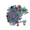
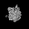
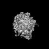
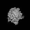
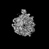
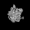
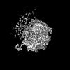
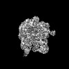
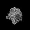
| Title | WT 12C IM fraction, B-b3 with RluB bound | |||||||||
 Components Components |
| |||||||||
 Keywords Keywords | RIBOSOME / 50S subunit / assembly intermediate / RNA-protein complex | |||||||||
| Function / homology |  Function and homology information Function and homology information23S rRNA pseudouridine2605 synthase / 23S rRNA pseudouridine(2605) synthase activity / rRNA pseudouridine synthase activity / enzyme-directed rRNA pseudouridine synthesis / transcriptional attenuation / endoribonuclease inhibitor activity / RNA-binding transcription regulator activity / negative regulation of cytoplasmic translation / translation repressor activity / mRNA regulatory element binding translation repressor activity ...23S rRNA pseudouridine2605 synthase / 23S rRNA pseudouridine(2605) synthase activity / rRNA pseudouridine synthase activity / enzyme-directed rRNA pseudouridine synthesis / transcriptional attenuation / endoribonuclease inhibitor activity / RNA-binding transcription regulator activity / negative regulation of cytoplasmic translation / translation repressor activity / mRNA regulatory element binding translation repressor activity / cytosolic ribosome assembly / ribosome assembly / assembly of large subunit precursor of preribosome / DNA-templated transcription termination / mRNA 5'-UTR binding / large ribosomal subunit / ribosomal large subunit assembly / large ribosomal subunit rRNA binding / cytosolic large ribosomal subunit / cytoplasmic translation / negative regulation of translation / rRNA binding / structural constituent of ribosome / ribosome / translation / response to antibiotic / negative regulation of DNA-templated transcription / mRNA binding / DNA binding / RNA binding / zinc ion binding / cytosol / cytoplasm Similarity search - Function | |||||||||
| Biological species |  | |||||||||
| Method | ELECTRON MICROSCOPY / single particle reconstruction / cryo EM / Resolution: 5.04 Å | |||||||||
 Authors Authors | Lee, J. / Sheng, K. / Williamson, J.R. | |||||||||
| Funding support |  United States, 2items United States, 2items
| |||||||||
 Citation Citation | Journal: To Be Published Title: 50S ribosome assembly intermediates at low temperature reveal bound RluB Authors: Lee, J. / Sheng, K. / Williamson, J.R. / Gebert, L. | |||||||||
| History |
|
- Structure visualization
| Structure viewer | Molecule: MolmilJmol/JSmol |
|---|
- Downloads & links
-Download
| PDBx/mmCIF format | 9cl9.cif.gz | 1.3 MB | Display | PDBx/mmCIF format |
|---|---|---|---|---|
| PDB format | pdb9cl9.ent.gz | Display | PDB format | |
| PDBx/mmJSON format | 9cl9.json.gz | Tree view | PDBx/mmJSON format | |
| Others |  Other downloads Other downloads |
-Validation report
| Arichive directory | https://data.pdbj.org/pub/pdb/validation_reports/cl/9cl9ftp://data.pdbj.org/pub/pdb/validation_reports/cl/9cl9 | HTTPS FTP |
|---|
-Related structure data
| Related structure data |  45666MC C: citing same article ( M: map data used to model this data |
|---|---|
| Similar structure data |


-Links
 PDBj
PDBj


- Assembly
| Deposited unit | 
|
|---|---|
| 1 |
|
-Components
-Large ribosomal subunit protein ... , 10 types, 10 molecules QRT2DEJSUY
| #1: Protein | Mass: 13396.828 Da / Num. of mol.: 1 / Source method: isolated from a natural source / Source: (natural) |
|---|---|
| #2: Protein | Mass: 11586.374 Da / Num. of mol.: 1 / Source method: isolated from a natural source / Source: (natural) |
| #3: Protein | Mass: 10144.924 Da / Num. of mol.: 1 / Source method: isolated from a natural source / Source: (natural) |
| #4: Protein/peptide | Mass: 4290.116 Da / Num. of mol.: 1 / Source method: isolated from a natural source / Source: (natural) |
| #7: Protein | Mass: 22277.535 Da / Num. of mol.: 1 / Source method: isolated from a natural source / Source: (natural) |
| #8: Protein | Mass: 22121.566 Da / Num. of mol.: 1 / Source method: isolated from a natural source / Source: (natural) |
| #9: Protein | Mass: 16050.606 Da / Num. of mol.: 1 / Source method: isolated from a natural source / Source: (natural) |
| #10: Protein | Mass: 12253.359 Da / Num. of mol.: 1 / Source method: isolated from a natural source / Source: (natural) |
| #11: Protein | Mass: 11078.874 Da / Num. of mol.: 1 / Source method: isolated from a natural source / Source: (natural) |
| #12: Protein | Mass: 7087.256 Da / Num. of mol.: 1 / Source method: isolated from a natural source / Source: (natural) |
-Protein / RNA chain , 2 types, 2 molecules ACA
| #5: Protein | Mass: 32010.760 Da / Num. of mol.: 1 / Source method: isolated from a natural source / Source: (natural) References: UniProt: P37765, 23S rRNA pseudouridine2605 synthase |
|---|---|
| #6: RNA chain | Mass: 941625.438 Da / Num. of mol.: 1 / Source method: isolated from a natural source Details: Base U at position 2605 on 23S rRNA (Chain CA:1281) could also be PSU. Source: (natural) |
-Details
| Has ligand of interest | N |
|---|
-Experimental details
-Experiment
| Experiment | Method: ELECTRON MICROSCOPY |
|---|---|
| EM experiment | Aggregation state: PARTICLE / 3D reconstruction method: single particle reconstruction |
- Sample preparation
| Component | Name: WT 12C 50S ribosomal subunit assembly intermediate / Type: RIBOSOME / Entity ID: all / Source: NATURAL | ||||||||||||||||||||||||||||||
|---|---|---|---|---|---|---|---|---|---|---|---|---|---|---|---|---|---|---|---|---|---|---|---|---|---|---|---|---|---|---|---|
| Molecular weight | Experimental value: NO | ||||||||||||||||||||||||||||||
| Source (natural) | Organism: | ||||||||||||||||||||||||||||||
| Buffer solution | pH: 7.5 Details: Most of the sucrose was removed by spin concentration. | ||||||||||||||||||||||||||||||
| Buffer component |
| ||||||||||||||||||||||||||||||
| Specimen | Embedding applied: NO / Shadowing applied: NO / Staining applied: NO / Vitrification applied: YES | ||||||||||||||||||||||||||||||
| Specimen support | Grid material: COPPER / Grid type: Quantifoil R1.2/1.3 | ||||||||||||||||||||||||||||||
| Vitrification | Instrument: FEI VITROBOT MARK IV / Cryogen name: ETHANE / Humidity: 90 % / Chamber temperature: 298 K / Details: 3 microliter of the sample was added. |
- Electron microscopy imaging
| Experimental equipment |  Model: Talos Arctica / Image courtesy: FEI Company |
|---|---|
| Microscopy | Model: FEI TALOS ARCTICA Details: In order to account for highly preferred orientation of the specimen, data were acquired using tilts at -20 degrees. |
| Electron gun | Electron source:  FIELD EMISSION GUN / Accelerating voltage: 200 kV / Illumination mode: FLOOD BEAM FIELD EMISSION GUN / Accelerating voltage: 200 kV / Illumination mode: FLOOD BEAM |
| Electron lens | Mode: BRIGHT FIELD / Nominal magnification: 36000 X / Nominal defocus max: 2000 nm / Nominal defocus min: 1000 nm / C2 aperture diameter: 50 µm |
| Specimen holder | Cryogen: NITROGEN |
| Image recording | Average exposure time: 5 sec. / Electron dose: 50 e/Å2 / Detector mode: COUNTING / Film or detector model: GATAN K2 SUMMIT (4k x 4k) / Num. of grids imaged: 1 |
- Processing
| EM software |
| ||||||||||||||||||||||||||||||||||||||||||||||||||||||||||||||||||||||||||||||||||||||||||||||||||||||||
|---|---|---|---|---|---|---|---|---|---|---|---|---|---|---|---|---|---|---|---|---|---|---|---|---|---|---|---|---|---|---|---|---|---|---|---|---|---|---|---|---|---|---|---|---|---|---|---|---|---|---|---|---|---|---|---|---|---|---|---|---|---|---|---|---|---|---|---|---|---|---|---|---|---|---|---|---|---|---|---|---|---|---|---|---|---|---|---|---|---|---|---|---|---|---|---|---|---|---|---|---|---|---|---|---|---|
| CTF correction | Details: CTF correction were performed by Patch CTF Estimation of cryoSPARC Type: NONE | ||||||||||||||||||||||||||||||||||||||||||||||||||||||||||||||||||||||||||||||||||||||||||||||||||||||||
| 3D reconstruction | Resolution: 5.04 Å / Resolution method: FSC 0.143 CUT-OFF / Num. of particles: 22502 / Symmetry type: POINT | ||||||||||||||||||||||||||||||||||||||||||||||||||||||||||||||||||||||||||||||||||||||||||||||||||||||||
| Atomic model building | B value: 118.8 / Protocol: OTHER / Space: REAL / Target criteria: Comprehensive validation (cryo-EM) Details: The atomic model was fitted into a low resolution (5 angstrom) EM map; therefore the coordinates should only serve as a general visual representation and should not be looked into detail. ...Details: The atomic model was fitted into a low resolution (5 angstrom) EM map; therefore the coordinates should only serve as a general visual representation and should not be looked into detail. Initial docking of the atomic model was done in ChimeraX. Coot and Phenix were used for real-space refinements. Geometry minimization in Phenix was used to improve the RNA atomic model. | ||||||||||||||||||||||||||||||||||||||||||||||||||||||||||||||||||||||||||||||||||||||||||||||||||||||||
| Atomic model building | 3D fitting-ID: 1
|