Movie
Movie Controller


+ Open data

- Basic information
| Entry |  | |||||||||
|---|---|---|---|---|---|---|---|---|---|---|
| Title | WT 12C IM fraction, B-b3 | |||||||||
 Map data Map data | WT 12C IM fraction, B-b3 | |||||||||
 Sample Sample |
| |||||||||
 Keywords Keywords | 50S subunit / assembly intermediate / RNA-protein complex / ribosome | |||||||||
| Biological species |  | |||||||||
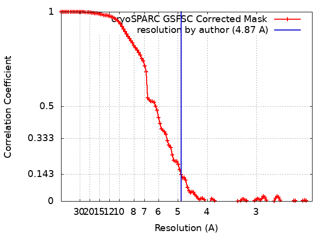
| Method | single particle reconstruction / cryo EM / Resolution: 4.87 Å | |||||||||
 Authors Authors | Lee J / Sheng K / Williamson JR | |||||||||
| Funding support |  United States, 2 items United States, 2 items
| |||||||||
 Citation Citation | Journal: To Be Published Title: 50S ribosome assembly intermediates at low temperature reveal bound RluB Authors: Lee J / Sheng K / Williamson JR / Gebert L | |||||||||
| History |
|
- Structure visualization
| Supplemental images |
|---|

- Downloads & links
-EMDB archive
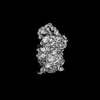
| Map data | emd_44834.map.gz | 136.5 MB |  EMDB map data format EMDB map data format | |
|---|---|---|---|---|
| Header (meta data) | emd-44834-v30.xmlemd-44834.xml | 14.8 KB 14.8 KB | Display Display | EMDB header |
| FSC (resolution estimation) | emd_44834_fsc.xml | 11.2 KB | Display | FSC data file |
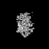
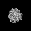
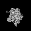
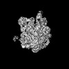
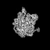
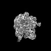
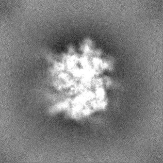
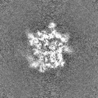
| Images |  emd_44834.png emd_44834.png | 74.1 KB | ||
| Filedesc metadata | emd-44834.cif.gz | 4.3 KB | ||
| Others | emd_44834_half_map_1.map.gzemd_44834_half_map_2.map.gz | 134.2 MB 134.2 MB | ||
| Archive directory |  http://ftp.pdbj.org/pub/emdb/structures/EMD-44834ftp://ftp.pdbj.org/pub/emdb/structures/EMD-44834 http://ftp.pdbj.org/pub/emdb/structures/EMD-44834ftp://ftp.pdbj.org/pub/emdb/structures/EMD-44834 | HTTPS FTP |
-Related structure data


-Links
| EMDB pages | EMDB (EBI/PDBe) / EMDataResource |
|---|
-Map
| File | Download / File: emd_44834.map.gz / Format: CCP4 / Size: 144.7 MB / Type: IMAGE STORED AS FLOATING POINT NUMBER (4 BYTES) | ||||||||||||||||||||||||||||||||||||
|---|---|---|---|---|---|---|---|---|---|---|---|---|---|---|---|---|---|---|---|---|---|---|---|---|---|---|---|---|---|---|---|---|---|---|---|---|---|
| Annotation | WT 12C IM fraction, B-b3 | ||||||||||||||||||||||||||||||||||||
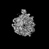
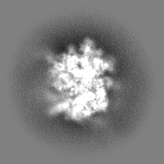
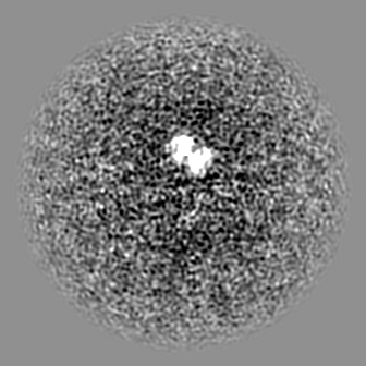
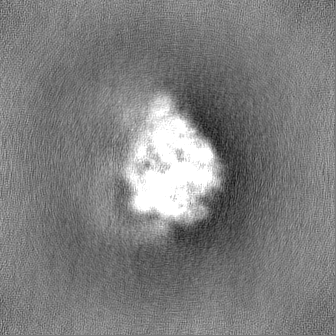
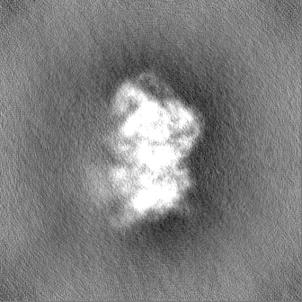
| Projections & slices | Image control
Images are generated by Spider. | ||||||||||||||||||||||||||||||||||||
| Voxel size | X=Y=Z: 1.15 Å | ||||||||||||||||||||||||||||||||||||
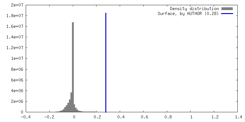
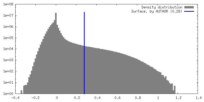
| Density |
| ||||||||||||||||||||||||||||||||||||
| Symmetry | Space group: 1 | ||||||||||||||||||||||||||||||||||||
| Details | EMDB XML:
|
 Z (Sec.)
 Y (Row.)
 X (Col.)


-Supplemental data
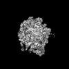
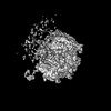
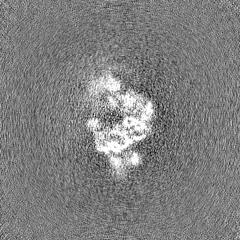
-Half map: WT 12C IM fraction, B-b3, half map A
| File | emd_44834_half_map_1.map | ||||||||||||
|---|---|---|---|---|---|---|---|---|---|---|---|---|---|
| Annotation | WT 12C IM fraction, B-b3, half map A | ||||||||||||
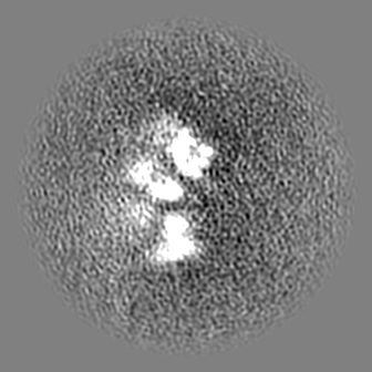
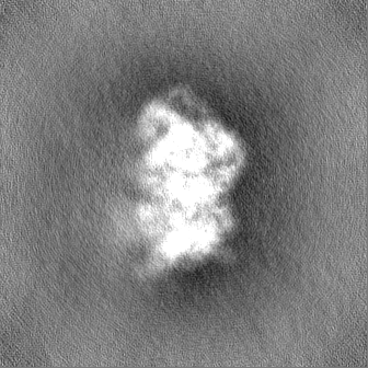
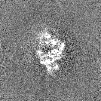
| Projections & Slices |
| ||||||||||||
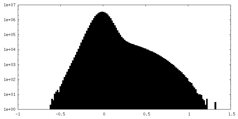
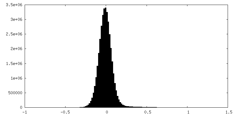
| Density Histograms |


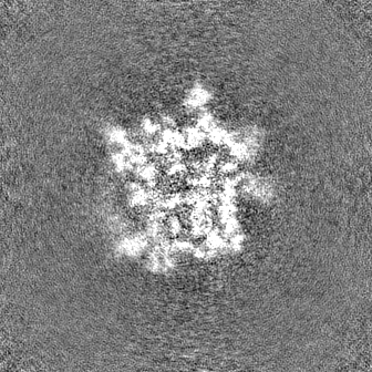

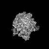
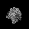
-Half map: WT 12C IM fraction, B-b3, half map B
| File | emd_44834_half_map_2.map | ||||||||||||
|---|---|---|---|---|---|---|---|---|---|---|---|---|---|
| Annotation | WT 12C IM fraction, B-b3, half map B | ||||||||||||
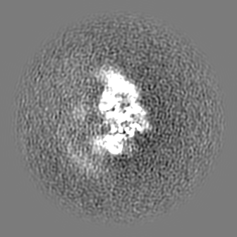
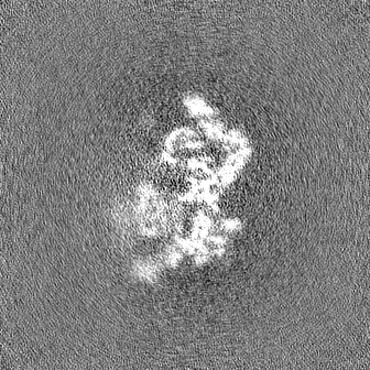
| Projections & Slices |
| ||||||||||||
| Density Histograms |


- Sample components
-Entire : WT 12C 50S ribosomal subunit assembly intermediate
| Entire | Name: WT 12C 50S ribosomal subunit assembly intermediate |
|---|---|
| Components |
|
-Supramolecule #1: WT 12C 50S ribosomal subunit assembly intermediate
| Supramolecule | Name: WT 12C 50S ribosomal subunit assembly intermediate / type: complex / ID: 1 / Parent: 0 |
|---|---|
| Source (natural) | Organism: |
-Experimental details
-Structure determination
| Method | cryo EM |
|---|---|
 Processing Processing | single particle reconstruction |
| Aggregation state | particle |
-Sample preparation
| Buffer | pH: 7.5 Component:
Details: Most of the sucrose was removed by spin concentration. | ||||||||||||||||||
|---|---|---|---|---|---|---|---|---|---|---|---|---|---|---|---|---|---|---|---|
| Grid | Model: Quantifoil R1.2/1.3 / Material: COPPER / Support film - Material: CARBON / Support film - topology: CONTINUOUS / Support film - Film thickness: 2 | ||||||||||||||||||
| Vitrification | Cryogen name: ETHANE / Chamber humidity: 90 % / Chamber temperature: 298 K / Instrument: FEI VITROBOT MARK IV / Details: 3 microliter of the sample was added.. |
- Electron microscopy
| Microscope | FEI TALOS ARCTICA |
|---|---|
| Details | In order to account for highly preferred orientation of the specimen, data were acquired using tilts at -20 degrees. |
| Image recording | Film or detector model: GATAN K2 SUMMIT (4k x 4k) / Detector mode: COUNTING / Number grids imaged: 1 / Average exposure time: 5.0 sec. / Average electron dose: 50.0 e/Å2 |
| Electron beam | Acceleration voltage: 200 kV / Electron source:  FIELD EMISSION GUN FIELD EMISSION GUN |
| Electron optics | C2 aperture diameter: 50.0 µm / Illumination mode: FLOOD BEAM / Imaging mode: BRIGHT FIELD / Nominal defocus max: 2.0 µm / Nominal defocus min: 1.0 µm / Nominal magnification: 36000 |
| Sample stage | Cooling holder cryogen: NITROGEN |
| Experimental equipment |  Model: Talos Arctica / Image courtesy: FEI Company |