 Movie
Movie Controller


+ Open data

- Basic information
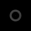
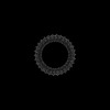
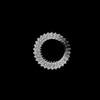
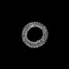
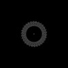
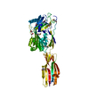
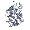
| Entry | Database: PDB / ID: 9ccq | ||||||||||||
|---|---|---|---|---|---|---|---|---|---|---|---|---|---|
| Title | Cryo-EM structure of the prepore-like EaCDCL short oligomer | ||||||||||||
 Components Components | Thiol-activated cytolysin family protein | ||||||||||||
 Keywords Keywords | TOXIN / pore-forming toxin / cholesterol-dependent cytolysin like / Elizabethkingia anophelis / MACPF / complement | ||||||||||||
| Function / homology | Thiol-activated cytolysin / Thiol-activated cytolysin superfamily / Thiol-activated cytolysin, alpha-beta domain superfamily / Thiol-activated cytolysin / cholesterol binding / Prokaryotic membrane lipoprotein lipid attachment site profile. / metal ion binding / Thiol-activated cytolysin family protein Function and homology information Function and homology information | ||||||||||||
| Biological species |  Elizabethkingia anophelis Ag1 (bacteria) Elizabethkingia anophelis Ag1 (bacteria) | ||||||||||||
| Method | ELECTRON MICROSCOPY / single particle reconstruction / cryo EM / Resolution: 3.13 Å | ||||||||||||
 Authors Authors | Johnstone, B.A. / Christie, M.P. / Morton, C.M. / Brown, H.G. / Hanssen, E. / Parker, M.W. | ||||||||||||
| Funding support |  Australia, 3items Australia, 3items
| ||||||||||||
 Citation Citation | Journal: Sci Adv / Year: 2025 Title: Structural basis for the pore-forming activity of a complement-like toxin. Authors: Bronte A Johnstone / Michelle P Christie / Riya Joseph / Craig J Morton / Hamish G Brown / Eric Hanssen / Tristan C Sanford / Hunter L Abrahamsen / Rodney K Tweten / Michael W Parker /  Abstract: Pore-forming proteins comprise a highly diverse group of proteins exemplified by the membrane attack complex/perforin (MACPF), cholesterol-dependent cytolysin (CDC), and gasdermin superfamilies, ...Pore-forming proteins comprise a highly diverse group of proteins exemplified by the membrane attack complex/perforin (MACPF), cholesterol-dependent cytolysin (CDC), and gasdermin superfamilies, which all form gigantic pores (>150 angstroms). A recently found family of pore-forming toxins, called CDC-like proteins (CDCLs), are wide-spread in gut microbes and are a prevalent means of antibacterial antagonism. However, the structural aspects of how CDCLs assemble a pore remain a mystery. Here, we report the crystal structure of a proteolytically activated CDCL and cryo-electron microscopy structures of a prepore-like intermediate and a transmembrane pore providing detailed snapshots across the entire pore-forming pathway. These studies reveal a sophisticated array of regulatory features to ensure productive pore formation, and, thus, CDCLs straddle the MACPF, CDC, and gasdermin lineages of the giant pore superfamilies. | ||||||||||||
| History |
|
- Structure visualization
| Structure viewer | Molecule: MolmilJmol/JSmol |
|---|
- Downloads & links
-Download
| PDBx/mmCIF format | 9ccq.cif.gz | 1.3 MB | Display | PDBx/mmCIF format |
|---|---|---|---|---|
| PDB format | pdb9ccq.ent.gz | Display | PDB format | |
| PDBx/mmJSON format | 9ccq.json.gz | Tree view | PDBx/mmJSON format | |
| Others |  Other downloads Other downloads |
-Validation report
| Arichive directory | https://data.pdbj.org/pub/pdb/validation_reports/cc/9ccqftp://data.pdbj.org/pub/pdb/validation_reports/cc/9ccq | HTTPS FTP |
|---|
-Related structure data
| Related structure data | 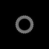 45453MC  8g33C 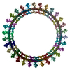 9ccpC C: citing same article ( M: map data used to model this data |
|---|---|
| Similar structure data |

-Links
 PDBj
PDBj


- Assembly
| Deposited unit | 
|
|---|---|
| 1 |
|
-Components
| #1: Protein | Mass: 39494.047 Da / Num. of mol.: 30 Source method: isolated from a genetically manipulated source Source: (gene. exp.) Elizabethkingia anophelis Ag1 (bacteria)Gene: JCR23_19030 / Production host: #2: Chemical | ChemComp-CA /   Mass: 40.078 Da / Num. of mol.: 30 / Source method: obtained synthetically / Formula: Ca Mass: 40.078 Da / Num. of mol.: 30 / Source method: obtained synthetically / Formula: CaHas ligand of interest | N | Has protein modification | N | |
|---|
-Experimental details
-Experiment
| Experiment | Method: ELECTRON MICROSCOPY |
|---|---|
| EM experiment | Aggregation state: PARTICLE / 3D reconstruction method: single particle reconstruction |
- Sample preparation
| Component | Name: EaCDCL pore embedded in POPC liposome / Type: COMPLEX / Entity ID: #1 / Source: RECOMBINANT |
|---|---|
| Molecular weight | Experimental value: NO |
| Source (natural) | Organism: Elizabethkingia anophelis Ag1 (bacteria) |
| Source (recombinant) | Organism: |
| Buffer solution | pH: 7.4 / Details: HBS pH 7.4 |
| Specimen | Embedding applied: NO / Shadowing applied: NO / Staining applied: NO / Vitrification applied: YES Details: Proteoliposome sample. Act-EaCDCLL + act-EaCDCLS (1:2 molar ratio) were added to liposomes to yield a final sample with a liposome concentration of 3.95 mM and a 1:500 protein:lipid molar ...Details: Proteoliposome sample. Act-EaCDCLL + act-EaCDCLS (1:2 molar ratio) were added to liposomes to yield a final sample with a liposome concentration of 3.95 mM and a 1:500 protein:lipid molar ratio. Sample was incubated at 37 degrees for 15 - 20 min before applying to grids. |
| Specimen support | Grid material: GOLD / Grid type: Quantifoil R2/2 |
| Vitrification | Instrument: FEI VITROBOT MARK IV / Cryogen name: ETHANE / Humidity: 100 % / Chamber temperature: 295 K |
- Electron microscopy imaging
| Experimental equipment |  Model: Titan Krios / Image courtesy: FEI Company |
|---|---|
| Microscopy | Model: FEI TITAN KRIOS |
| Electron gun | Electron source:  FIELD EMISSION GUN / Accelerating voltage: 300 kV / Illumination mode: FLOOD BEAM FIELD EMISSION GUN / Accelerating voltage: 300 kV / Illumination mode: FLOOD BEAM |
| Electron lens | Mode: BRIGHT FIELD / Nominal magnification: 64000 X / Nominal defocus max: 2000 nm / Nominal defocus min: 800 nm / Cs: 2.7 mm |
| Specimen holder | Specimen holder model: FEI TITAN KRIOS AUTOGRID HOLDER |
| Image recording | Electron dose: 50 e/Å2 / Film or detector model: GATAN K3 (6k x 4k) / Num. of grids imaged: 1 / Num. of real images: 15971 |
| EM imaging optics | Energyfilter slit width: 20 eV |
- Processing
| EM software | Name: PHENIX / Version: 1.20.1_4487: / Category: model refinement | ||||||||||||||||||||||||
|---|---|---|---|---|---|---|---|---|---|---|---|---|---|---|---|---|---|---|---|---|---|---|---|---|---|
| CTF correction | Type: PHASE FLIPPING AND AMPLITUDE CORRECTION | ||||||||||||||||||||||||
| Particle selection | Num. of particles selected: 559234 | ||||||||||||||||||||||||
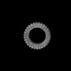
| Symmetry | Point symmetry: C30 (30 fold cyclic) | ||||||||||||||||||||||||
| 3D reconstruction | Resolution: 3.13 Å / Resolution method: FSC 0.143 CUT-OFF / Num. of particles: 91117 / Symmetry type: POINT | ||||||||||||||||||||||||
| Atomic model building | Protocol: RIGID BODY FIT | ||||||||||||||||||||||||
| Atomic model building | PDB-ID: 8G32 Pdb chain-ID: A / Accession code: 8G32 / Source name: PDB / Type: experimental model | ||||||||||||||||||||||||
| Refine LS restraints |
|
