Movie
Movie Controller


+ Open data

- Basic information
| Entry |  | ||||||||||||
|---|---|---|---|---|---|---|---|---|---|---|---|---|---|
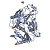
| Title | Cryo-EM structure of the prepore-like EaCDCL short oligomer | ||||||||||||
 Map data Map data | Sharpened map | ||||||||||||
 Sample Sample |
| ||||||||||||
 Keywords Keywords | pore-forming toxin / cholesterol-dependent cytolysin like / Elizabethkingia anophelis / MACPF / complement / TOXIN | ||||||||||||
| Function / homology | Thiol-activated cytolysin / Thiol-activated cytolysin superfamily / Thiol-activated cytolysin, alpha-beta domain superfamily / Thiol-activated cytolysin / cholesterol binding / Prokaryotic membrane lipoprotein lipid attachment site profile. / metal ion binding / Thiol-activated cytolysin family protein Function and homology information Function and homology information | ||||||||||||
| Biological species |  Elizabethkingia anophelis Ag1 (bacteria) Elizabethkingia anophelis Ag1 (bacteria) | ||||||||||||
| Method | single particle reconstruction / cryo EM / Resolution: 3.13 Å | ||||||||||||
 Authors Authors | Johnstone BA / Christie MP / Morton CM / Brown HG / Hanssen E / Parker MW | ||||||||||||
| Funding support |  Australia, 3 items Australia, 3 items
| ||||||||||||
 Citation Citation | Journal: Sci Adv / Year: 2025 Title: Structural basis for the pore-forming activity of a complement-like toxin. Authors: Bronte A Johnstone / Michelle P Christie / Riya Joseph / Craig J Morton / Hamish G Brown / Eric Hanssen / Tristan C Sanford / Hunter L Abrahamsen / Rodney K Tweten / Michael W Parker /  Abstract: Pore-forming proteins comprise a highly diverse group of proteins exemplified by the membrane attack complex/perforin (MACPF), cholesterol-dependent cytolysin (CDC), and gasdermin superfamilies, ...Pore-forming proteins comprise a highly diverse group of proteins exemplified by the membrane attack complex/perforin (MACPF), cholesterol-dependent cytolysin (CDC), and gasdermin superfamilies, which all form gigantic pores (>150 angstroms). A recently found family of pore-forming toxins, called CDC-like proteins (CDCLs), are wide-spread in gut microbes and are a prevalent means of antibacterial antagonism. However, the structural aspects of how CDCLs assemble a pore remain a mystery. Here, we report the crystal structure of a proteolytically activated CDCL and cryo-electron microscopy structures of a prepore-like intermediate and a transmembrane pore providing detailed snapshots across the entire pore-forming pathway. These studies reveal a sophisticated array of regulatory features to ensure productive pore formation, and, thus, CDCLs straddle the MACPF, CDC, and gasdermin lineages of the giant pore superfamilies. | ||||||||||||
| History |
|
- Structure visualization
| Supplemental images |
|---|

- Downloads & links
-EMDB archive
| Map data | emd_45453.map.gz | 483.7 MB | EMDB map data format | |
|---|---|---|---|---|
| Header (meta data) | emd-45453-v30.xmlemd-45453.xml | 19.8 KB 19.8 KB | Display Display | EMDB header |
| FSC (resolution estimation) | emd_45453_fsc.xml | 16.9 KB | Display | FSC data file |
| Images |  emd_45453.png emd_45453.png | 112.3 KB | ||
| Masks | emd_45453_msk_1.map | 512 MB | Mask map | |
| Filedesc metadata | emd-45453.cif.gz | 6.6 KB | ||
| Others | emd_45453_additional_1.map.gzemd_45453_half_map_1.map.gzemd_45453_half_map_2.map.gz | 254.1 MB 474 MB 474.1 MB | ||
| Archive directory |  http://ftp.pdbj.org/pub/emdb/structures/EMD-45453ftp://ftp.pdbj.org/pub/emdb/structures/EMD-45453 http://ftp.pdbj.org/pub/emdb/structures/EMD-45453ftp://ftp.pdbj.org/pub/emdb/structures/EMD-45453 | HTTPS FTP |
-Related structure data
| Related structure data | 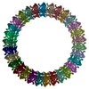 9ccqMC 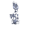 8g33C 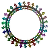 9ccpC M: atomic model generated by this map C: citing same article ( |
|---|---|
| Similar structure data |

-Links
| EMDB pages | EMDB (EBI/PDBe) / EMDataResource |
|---|---|
| Related items in Molecule of the Month |


-Map
| File | Download / File: emd_45453.map.gz / Format: CCP4 / Size: 512 MB / Type: IMAGE STORED AS FLOATING POINT NUMBER (4 BYTES) | ||||||||||||||||||||||||||||||||||||
|---|---|---|---|---|---|---|---|---|---|---|---|---|---|---|---|---|---|---|---|---|---|---|---|---|---|---|---|---|---|---|---|---|---|---|---|---|---|
| Annotation | Sharpened map | ||||||||||||||||||||||||||||||||||||
| Projections & slices | Image control
Images are generated by Spider. | ||||||||||||||||||||||||||||||||||||
| Voxel size | X=Y=Z: 1.32 Å | ||||||||||||||||||||||||||||||||||||
| Density |
| ||||||||||||||||||||||||||||||||||||
| Symmetry | Space group: 1 | ||||||||||||||||||||||||||||||||||||
| Details | EMDB XML:
|
 Z (Sec.)
 Y (Row.)
 X (Col.)


-Supplemental data
-Mask #1
| File | emd_45453_msk_1.map | ||||||||||||
|---|---|---|---|---|---|---|---|---|---|---|---|---|---|
| Projections & Slices |
| ||||||||||||
| Density Histograms |


-Additional map: Raw, unsharpened map
| File | emd_45453_additional_1.map | ||||||||||||
|---|---|---|---|---|---|---|---|---|---|---|---|---|---|
| Annotation | Raw, unsharpened map | ||||||||||||
| Projections & Slices |
| ||||||||||||
| Density Histograms |


-Half map: Half map 1
| File | emd_45453_half_map_1.map | ||||||||||||
|---|---|---|---|---|---|---|---|---|---|---|---|---|---|
| Annotation | Half map 1 | ||||||||||||
| Projections & Slices |
| ||||||||||||
| Density Histograms |


-Half map: Half map 2
| File | emd_45453_half_map_2.map | ||||||||||||
|---|---|---|---|---|---|---|---|---|---|---|---|---|---|
| Annotation | Half map 2 | ||||||||||||
| Projections & Slices |
| ||||||||||||
| Density Histograms |


- Sample components
-Entire : EaCDCL pore embedded in POPC liposome
| Entire | Name: EaCDCL pore embedded in POPC liposome |
|---|---|
| Components |
|
-Supramolecule #1: EaCDCL pore embedded in POPC liposome
| Supramolecule | Name: EaCDCL pore embedded in POPC liposome / type: complex / ID: 1 / Parent: 0 / Macromolecule list: #1 |
|---|---|
| Source (natural) | Organism: Elizabethkingia anophelis Ag1 (bacteria) |
-Macromolecule #1: Thiol-activated cytolysin family protein
| Macromolecule | Name: Thiol-activated cytolysin family protein / type: protein_or_peptide / ID: 1 / Number of copies: 30 / Enantiomer: LEVO |
|---|---|
| Source (natural) | Organism: Elizabethkingia anophelis Ag1 (bacteria) |
| Molecular weight | Theoretical: 39.494047 KDa |
| Recombinant expression | Organism: |
| Sequence | String: GSHMRQDSEV NPLQVQNSSK VLNPNVTLPA NNLLYDEFFV SKESKLIEDS RNNKRKTSKI ASLNPYASTK AVLTTTSSTL TSDQIVVTV PQKTFIGGVY NSTTLDNLDY TPISYPLDPI TVSYSFPSDF IVDTIERPSL SSMRASVFKA MRAANFSGEQ S LAFDYNIK ...String: GSHMRQDSEV NPLQVQNSSK VLNPNVTLPA NNLLYDEFFV SKESKLIEDS RNNKRKTSKI ASLNPYASTK AVLTTTSSTL TSDQIVVTV PQKTFIGGVY NSTTLDNLDY TPISYPLDPI TVSYSFPSDF IVDTIERPSL SSMRASVFKA MRAANFSGEQ S LAFDYNIK QFSYYSELKI AFGSNVNIGK IFSIDISGSN NKIKRTTGVF AKFTQKNFTI DMDLPADGNI FKNNSDLALT NG KNPVYIS SVTYGRLGII SIESNASYNE VNFALKAALT AGIVNGSLNI DSNSKKILEE SDLSVYLVGG RGTDAVQVIK GFA GFSNFI VNGGQFTPEA PGVPIYFSAS HASDNSVYYT TFTIDK UniProtKB: Thiol-activated cytolysin family protein |
-Macromolecule #2: CALCIUM ION
| Macromolecule | Name: CALCIUM ION / type: ligand / ID: 2 / Number of copies: 30 / Formula: CA |
|---|---|
| Molecular weight | Theoretical: 40.078 Da |
-Experimental details
-Structure determination
| Method | cryo EM |
|---|---|
 Processing Processing | single particle reconstruction |
| Aggregation state | particle |
-Sample preparation
| Buffer | pH: 7.4 / Details: HBS pH 7.4 |
|---|---|
| Grid | Model: Quantifoil R2/2 / Material: GOLD / Support film - Material: CARBON / Support film - topology: CONTINUOUS / Support film - Film thickness: 2 |
| Vitrification | Cryogen name: ETHANE / Chamber humidity: 100 % / Chamber temperature: 295 K / Instrument: FEI VITROBOT MARK IV |
| Details | Proteoliposome sample. Act-EaCDCLL + act-EaCDCLS (1:2 molar ratio) were added to liposomes to yield a final sample with a liposome concentration of 3.95 mM and a 1:500 protein:lipid molar ratio. Sample was incubated at 37 degrees for 15 - 20 min before applying to grids. |
- Electron microscopy
| Microscope | FEI TITAN KRIOS |
|---|---|
| Specialist optics | Energy filter - Slit width: 20 eV |
| Image recording | Film or detector model: GATAN K3 (6k x 4k) / Number grids imaged: 1 / Number real images: 15971 / Average electron dose: 50.0 e/Å2 |
| Electron beam | Acceleration voltage: 300 kV / Electron source:  FIELD EMISSION GUN FIELD EMISSION GUN |
| Electron optics | Illumination mode: FLOOD BEAM / Imaging mode: BRIGHT FIELD / Cs: 2.7 mm / Nominal defocus max: 2.0 µm / Nominal defocus min: 0.8 µm / Nominal magnification: 64000 |
| Sample stage | Specimen holder model: FEI TITAN KRIOS AUTOGRID HOLDER |
| Experimental equipment |  Model: Titan Krios / Image courtesy: FEI Company |