 Movie
Movie Controller


+ Open data

- Basic information
| Entry |  | ||||||||||||
|---|---|---|---|---|---|---|---|---|---|---|---|---|---|
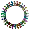
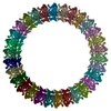
| Title | Double-stacked pore and prepore-like complex (C1 symmetry) | ||||||||||||
 Map data Map data | Sharpened map | ||||||||||||
 Sample Sample |
| ||||||||||||
 Keywords Keywords | pore-forming toxin / cholesterol-dependent cytolysin like / Elizabethkingia anophelis / MACPF / complement / TOXIN | ||||||||||||
| Function / homology | Thiol-activated cytolysin / Thiol-activated cytolysin superfamily / Thiol-activated cytolysin, alpha-beta domain superfamily / Thiol-activated cytolysin / cholesterol binding / Prokaryotic membrane lipoprotein lipid attachment site profile. / metal ion binding / Hemolysin / Thiol-activated cytolysin family protein Function and homology information Function and homology information | ||||||||||||
| Biological species |  Elizabethkingia anophelis Ag1 (bacteria) Elizabethkingia anophelis Ag1 (bacteria) | ||||||||||||
| Method | single particle reconstruction / cryo EM / Resolution: 3.35 Å | ||||||||||||
 Authors Authors | Johnstone BA / Christie MP / Morton CJ / Brown HG / Hanssen E / Parker MW | ||||||||||||
| Funding support |  Australia, 3 items Australia, 3 items
| ||||||||||||
 Citation Citation | Journal: Sci Adv / Year: 2025 Title: Structural basis for the pore-forming activity of a complement-like toxin. Authors: Bronte A Johnstone / Michelle P Christie / Riya Joseph / Craig J Morton / Hamish G Brown / Eric Hanssen / Tristan C Sanford / Hunter L Abrahamsen / Rodney K Tweten / Michael W Parker /  Abstract: Pore-forming proteins comprise a highly diverse group of proteins exemplified by the membrane attack complex/perforin (MACPF), cholesterol-dependent cytolysin (CDC), and gasdermin superfamilies, ...Pore-forming proteins comprise a highly diverse group of proteins exemplified by the membrane attack complex/perforin (MACPF), cholesterol-dependent cytolysin (CDC), and gasdermin superfamilies, which all form gigantic pores (>150 angstroms). A recently found family of pore-forming toxins, called CDC-like proteins (CDCLs), are wide-spread in gut microbes and are a prevalent means of antibacterial antagonism. However, the structural aspects of how CDCLs assemble a pore remain a mystery. Here, we report the crystal structure of a proteolytically activated CDCL and cryo-electron microscopy structures of a prepore-like intermediate and a transmembrane pore providing detailed snapshots across the entire pore-forming pathway. These studies reveal a sophisticated array of regulatory features to ensure productive pore formation, and, thus, CDCLs straddle the MACPF, CDC, and gasdermin lineages of the giant pore superfamilies. | ||||||||||||
| History |
|
- Structure visualization
| Supplemental images |
|---|

- Downloads & links
-EMDB archive
| Map data | emd_45448.map.gz | 483.8 MB | EMDB map data format | |
|---|---|---|---|---|
| Header (meta data) | emd-45448-v30.xmlemd-45448.xml | 20 KB 20 KB | Display Display | EMDB header |
| FSC (resolution estimation) | emd_45448_fsc.xml | 16.8 KB | Display | FSC data file |
| Images |  emd_45448.png emd_45448.png | 153.6 KB | ||
| Masks | emd_45448_msk_1.map | 512 MB | Mask map | |
| Filedesc metadata | emd-45448.cif.gz | 5.9 KB | ||
| Others | emd_45448_additional_1.map.gzemd_45448_half_map_1.map.gzemd_45448_half_map_2.map.gz | 256.9 MB 475.3 MB 475.3 MB | ||
| Archive directory |  http://ftp.pdbj.org/pub/emdb/structures/EMD-45448ftp://ftp.pdbj.org/pub/emdb/structures/EMD-45448 http://ftp.pdbj.org/pub/emdb/structures/EMD-45448ftp://ftp.pdbj.org/pub/emdb/structures/EMD-45448 | HTTPS FTP |
-Related structure data

-Links
| EMDB pages | EMDB (EBI/PDBe) / EMDataResource |
|---|---|
| Related items in Molecule of the Month |


-Map
| File | Download / File: emd_45448.map.gz / Format: CCP4 / Size: 512 MB / Type: IMAGE STORED AS FLOATING POINT NUMBER (4 BYTES) | ||||||||||||||||||||||||||||||||||||
|---|---|---|---|---|---|---|---|---|---|---|---|---|---|---|---|---|---|---|---|---|---|---|---|---|---|---|---|---|---|---|---|---|---|---|---|---|---|
| Annotation | Sharpened map | ||||||||||||||||||||||||||||||||||||
| Projections & slices | Image control
Images are generated by Spider. | ||||||||||||||||||||||||||||||||||||
| Voxel size | X=Y=Z: 1.32 Å | ||||||||||||||||||||||||||||||||||||
| Density |
| ||||||||||||||||||||||||||||||||||||
| Symmetry | Space group: 1 | ||||||||||||||||||||||||||||||||||||
| Details | EMDB XML:
|
 Z (Sec.)
 Y (Row.)
 X (Col.)


-Supplemental data
-Mask #1
| File | emd_45448_msk_1.map | ||||||||||||
|---|---|---|---|---|---|---|---|---|---|---|---|---|---|
| Projections & Slices |
| ||||||||||||
| Density Histograms |


-Additional map: Raw, unsharpened map
| File | emd_45448_additional_1.map | ||||||||||||
|---|---|---|---|---|---|---|---|---|---|---|---|---|---|
| Annotation | Raw, unsharpened map | ||||||||||||
| Projections & Slices |
| ||||||||||||
| Density Histograms |


-Half map: #2
| File | emd_45448_half_map_1.map | ||||||||||||
|---|---|---|---|---|---|---|---|---|---|---|---|---|---|
| Projections & Slices |
| ||||||||||||
| Density Histograms |


-Half map: #1
| File | emd_45448_half_map_2.map | ||||||||||||
|---|---|---|---|---|---|---|---|---|---|---|---|---|---|
| Projections & Slices |
| ||||||||||||
| Density Histograms |


- Sample components
-Entire : EaCDCL pore embedded in POPC liposome
| Entire | Name: EaCDCL pore embedded in POPC liposome |
|---|---|
| Components |
|
-Supramolecule #1: EaCDCL pore embedded in POPC liposome
| Supramolecule | Name: EaCDCL pore embedded in POPC liposome / type: complex / ID: 1 / Parent: 0 / Macromolecule list: all |
|---|---|
| Source (natural) | Organism: Elizabethkingia anophelis Ag1 (bacteria) |
-Macromolecule #1: EaCDCL short
| Macromolecule | Name: EaCDCL short / type: protein_or_peptide / ID: 1 / Enantiomer: LEVO |
|---|---|
| Source (natural) | Organism: Elizabethkingia anophelis Ag1 (bacteria) |
| Recombinant expression | Organism: |
| Sequence | String: GSHMRQDSEV NPLQVQNSSK VLNPNVTLPA NNLLYDEFFV SKESKLIEDS RNNKRKTSKI ASLNPYASTK AVLTTTSSTL TSDQIVVTVP QKTFIGGVYN STTLDNLDYT PISYPLDPIT VSYSFPSDFI VDTIERPSLS SMRASVFKAM RAANFSGEQS LAFDYNIKQF ...String: GSHMRQDSEV NPLQVQNSSK VLNPNVTLPA NNLLYDEFFV SKESKLIEDS RNNKRKTSKI ASLNPYASTK AVLTTTSSTL TSDQIVVTVP QKTFIGGVYN STTLDNLDYT PISYPLDPIT VSYSFPSDFI VDTIERPSLS SMRASVFKAM RAANFSGEQS LAFDYNIKQF SYYSELKIAF GSNVNIGKIF SIDISGSNNK IKRTTGVFAK FTQKNFTIDM DLPADGNIFK NNSDLALTNG KNPVYISSVT YGRLGIISIE SNASYNEVNF ALKAALTAGI VNGSLNIDSN SKKILEESDL SVYLVGGRGT DAVQVIKGFA GFSNFIVNGG QFTPEAPGVP IYFSASHASD NSVYYTTFTI DK UniProtKB: Thiol-activated cytolysin family protein |
-Macromolecule #2: EaCDCL long
| Macromolecule | Name: EaCDCL long / type: protein_or_peptide / ID: 2 / Enantiomer: LEVO |
|---|---|
| Source (natural) | Organism: Elizabethkingia anophelis Ag1 (bacteria) |
| Recombinant expression | Organism: |
| Sequence | String: GSHMTDNLPD RSSVTNNMAR VELPVINITS FGTKPSFLNI QKSASTKSLN LIAENSGDTE TKEFESSESV VLNHLNRYVF PGSLLMGNSI QDLNYKPVFA SLNPITVSLS IPAINQNTAI TITNPSLSAT RAAVYNYLKT ADFTQNGQLS YSIQQFSSYD ELKVAFGSNV ...String: GSHMTDNLPD RSSVTNNMAR VELPVINITS FGTKPSFLNI QKSASTKSLN LIAENSGDTE TKEFESSESV VLNHLNRYVF PGSLLMGNSI QDLNYKPVFA SLNPITVSLS IPAINQNTAI TITNPSLSAT RAAVYNYLKT ADFTQNGQLS YSIQQFSSYD ELKVAFGSNV NSRNLFGKNS SSTNVEEGMV ARQSGFYVKF YQTSFTLDMD VPNGSLVKDN NFDSEGIEPV YVSSISYGRM GILAIETNEK AEDAKRIINE TFNKLFYKKQ TNFSQEEKSF IEGADFNLYL VGGDGSTASQ SFKGYEAFVN HVSQGTFSKD QPGVPIFCSY SYLKDNSPVK TKFKFDIKRP PLYVKLVKEN MKDINFNDPD GGIYDNKKEA ILKIYFYKNR SLVPTLPNPY INFKIREKKK KWQSIAPVYY SSLDQVPFNI SERILTKQNT LQNIFATIQT QDNTEFSLIS RIIRGGGRQN PVPAGFRAIE INDYELVEDS NYIIIKD UniProtKB: Hemolysin |
-Experimental details
-Structure determination
| Method | cryo EM |
|---|---|
 Processing Processing | single particle reconstruction |
| Aggregation state | particle |
-Sample preparation
| Buffer | pH: 7.4 / Details: HBS pH 7.4 |
|---|---|
| Grid | Model: Quantifoil R2/2 / Material: GOLD / Support film - Material: CARBON / Support film - topology: CONTINUOUS / Support film - Film thickness: 2 |
| Vitrification | Cryogen name: ETHANE / Chamber humidity: 100 % / Chamber temperature: 295 K / Instrument: FEI VITROBOT MARK IV |
| Details | Proteoliposome sample. Act-EaCDCLL + act-EaCDCLS (1:2 molar ratio) were added to liposomes to yield a final sample with a liposome concentration of 3.95 mM and a 1:500 protein:lipid molar ratio. Sample was incubated at 37 degrees for 15 - 20 min before applying to grids. |
- Electron microscopy
| Microscope | FEI TITAN KRIOS |
|---|---|
| Specialist optics | Energy filter - Slit width: 20 eV |
| Image recording | Film or detector model: GATAN K3 (6k x 4k) / Number grids imaged: 1 / Number real images: 15971 / Average electron dose: 50.0 e/Å2 |
| Electron beam | Acceleration voltage: 300 kV / Electron source:  FIELD EMISSION GUN FIELD EMISSION GUN |
| Electron optics | Illumination mode: FLOOD BEAM / Imaging mode: BRIGHT FIELD / Cs: 2.7 mm / Nominal defocus max: 2.0 µm / Nominal defocus min: 0.8 µm / Nominal magnification: 64000 |
| Sample stage | Specimen holder model: FEI TITAN KRIOS AUTOGRID HOLDER |
| Experimental equipment |  Model: Titan Krios / Image courtesy: FEI Company |

