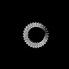
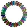
Journal: Sci Adv / Year: 2025 Title: Structural basis for the pore-forming activity of a complement-like toxin. Authors: Bronte A Johnstone / Michelle P Christie / Riya Joseph / Craig J Morton / Hamish G Brown / Eric Hanssen / Tristan C Sanford / Hunter L Abrahamsen / Rodney K Tweten / Michael W Parker / Abstract: Pore-forming proteins comprise a highly diverse group of proteins exemplified by the membrane attack complex/perforin (MACPF), cholesterol-dependent cytolysin (CDC), and gasdermin superfamilies, ...Pore-forming proteins comprise a highly diverse group of proteins exemplified by the membrane attack complex/perforin (MACPF), cholesterol-dependent cytolysin (CDC), and gasdermin superfamilies, which all form gigantic pores (>150 angstroms). A recently found family of pore-forming toxins, called CDC-like proteins (CDCLs), are wide-spread in gut microbes and are a prevalent means of antibacterial antagonism. However, the structural aspects of how CDCLs assemble a pore remain a mystery. Here, we report the crystal structure of a proteolytically activated CDCL and cryo-electron microscopy structures of a prepore-like intermediate and a transmembrane pore providing detailed snapshots across the entire pore-forming pathway. These studies reveal a sophisticated array of regulatory features to ensure productive pore formation, and, thus, CDCLs straddle the MACPF, CDC, and gasdermin lineages of the giant pore superfamilies.
In the structure databanks used in Yorodumi, some data are registered as the other names, "COVID-19 virus" and "2019-nCoV". Here are the details of the virus and the list of structure data.
Jan 31, 2019. EMDB accession codes are about to change! (news from PDBe EMDB page)
EMDB accession codes are about to change! (news from PDBe EMDB page)
The allocation of 4 digits for EMDB accession codes will soon come to an end. Whilst these codes will remain in use, new EMDB accession codes will include an additional digit and will expand incrementally as the available range of codes is exhausted. The current 4-digit format prefixed with “EMD-” (i.e. EMD-XXXX) will advance to a 5-digit format (i.e. EMD-XXXXX), and so on. It is currently estimated that the 4-digit codes will be depleted around Spring 2019, at which point the 5-digit format will come into force.
The EM Navigator/Yorodumi systems omit the EMD- prefix.
Related info.:Q: What is EMD? / ID/Accession-code notation in Yorodumi/EM Navigator
Yorodumi is a browser for structure data from EMDB, PDB, SASBDB, etc.
This page is also the successor to EM Navigator detail page, and also detail information page/front-end page for Omokage search.
The word "yorodu" (or yorozu) is an old Japanese word meaning "ten thousand". "mi" (miru) is to see.
Related info.:EMDB / PDB / SASBDB / Comparison of 3 databanks / Yorodumi Search / Aug 31, 2016. New EM Navigator & Yorodumi / Yorodumi Papers / Jmol/JSmol / Function and homology information / Changes in new EM Navigator and Yorodumi
 Movie
Movie Controller


 Open data
 Basic information
 Components
 Keywords
 Function and homology information
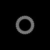
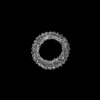
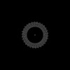
 Elizabethkingia anophelis Ag1 (bacteria)
 X-RAY DIFFRACTION /
  Authors
 Australia, 2items
  Citation
 Structure visualization
 Downloads & links
 Other downloads
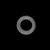

 PDBj
PDBj


 Assembly
 Mass: 18.015 Da / Num. of mol.: 45 / Source method: isolated from a natural source / Formula: H2O
 Sample preparation
 Processing