 Movie
Movie Controller


+ Open data

- Basic information
| Entry | Database: PDB / ID: 9bw6 | ||||||
|---|---|---|---|---|---|---|---|
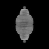
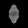
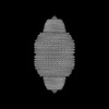
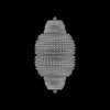
| Title | Human Vault Cage in complex with PARP4 | ||||||
 Components Components |
| ||||||
 Keywords Keywords | PROTEIN TRANSPORT / Vault / Vault Cage / MVP / Major Vault Protein / SPFH / PARP4 / Poly(ADP-ribose)Polymerase 4 / MINT / Poly(ADP-ribose)Polymerase / Ribonucleoprotein / Megadalton complex / TEP1 / vault RNA | ||||||
| Function / homology |  Function and homology information Function and homology informationprotein activation cascade / Maturation of nucleoprotein / Nicotinate metabolism / ERBB signaling pathway / Maturation of nucleoprotein / NAD+-protein-aspartate ADP-ribosyltransferase activity / NAD+-protein-glutamate ADP-ribosyltransferase activity / NAD+-protein mono-ADP-ribosyltransferase activity / Transferases; Glycosyltransferases; Pentosyltransferases / negative regulation of epidermal growth factor receptor signaling pathway ...protein activation cascade / Maturation of nucleoprotein / Nicotinate metabolism / ERBB signaling pathway / Maturation of nucleoprotein / NAD+-protein-aspartate ADP-ribosyltransferase activity / NAD+-protein-glutamate ADP-ribosyltransferase activity / NAD+-protein mono-ADP-ribosyltransferase activity / Transferases; Glycosyltransferases; Pentosyltransferases / negative regulation of epidermal growth factor receptor signaling pathway / NAD+ poly-ADP-ribosyltransferase activity / nuclear pore / mRNA transport / nucleotidyltransferase activity / protein modification process / spindle microtubule / protein transport / secretory granule lumen / protein phosphatase binding / ficolin-1-rich granule lumen / cytoskeleton / cell population proliferation / intracellular signal transduction / response to xenobiotic stimulus / ribonucleoprotein complex / inflammatory response / DNA repair / DNA damage response / Neutrophil degranulation / protein kinase binding / perinuclear region of cytoplasm / enzyme binding / DNA binding / extracellular exosome / extracellular region / nucleoplasm / membrane / identical protein binding / nucleus / cytoplasm / cytosol Similarity search - Function | ||||||
| Biological species |  Homo sapiens (human) Homo sapiens (human) | ||||||
| Method | ELECTRON MICROSCOPY / single particle reconstruction / cryo EM / Resolution: 2.9 Å | ||||||
 Authors Authors | Lodwick, J.E. / Zhao, M. | ||||||
| Funding support |  United States, 1items United States, 1items
| ||||||
 Citation Citation | Journal: Nat Commun / Year: 2025 Title: Structural insights into the roles of PARP4 and NAD binding in the human vault cage. Authors: Jane E Lodwick / Rong Shen / Satchal Erramilli / Yuan Xie / Karolina Roganowicz / Simone Ritchey / Anthony A Kossiakoff / Minglei Zhao /  Abstract: Vault is a massive ribonucleoprotein complex found across Eukaryota. The major vault protein (MVP) oligomerizes into an ovular cage, which contains several minor vault components (MVCs) and is ...Vault is a massive ribonucleoprotein complex found across Eukaryota. The major vault protein (MVP) oligomerizes into an ovular cage, which contains several minor vault components (MVCs) and is thought to transport transiently bound "cargo" molecules. Vertebrate vaults house a poly (ADP-ribose) polymerase (known as PARP4 in humans), which is the only MVC with known enzymatic activity. Despite being discovered decades ago, the molecular basis for PARP4's interaction with MVP remains unclear. In this study, we determined the structure of the human vault cage in complex with PARP4 and its enzymatic substrate NAD. The structures reveal atomic-level details of the protein-binding interface, as well as unexpected binding sites for NAD and related nucleotides within the interior of the vault cage. In addition, proteomics data show that human vaults purified from wild-type and PARP4-depleted cells interact with distinct subsets of proteins. Our results thereby support a model in which PARP4's specific incorporation into the vault cage helps to regulate vault's selection of cargo and its subcellular localization. Further, PARP4's proximity to MVP's NAD-binding sites could support its enzymatic function within the vault. #1: Journal: bioRxiv / Year: 2024Title: Structural Insights into the Roles of PARP4 and NAD in the Human Vault Cage. Abstract: Vault is a massive ribonucleoprotein complex found across Eukaryota. The major vault protein (MVP) oligomerizes into an ovular cage, which contains several minor vault components (MVCs) and is ...Vault is a massive ribonucleoprotein complex found across Eukaryota. The major vault protein (MVP) oligomerizes into an ovular cage, which contains several minor vault components (MVCs) and is thought to transport transiently bound "cargo" molecules. Vertebrate vaults house a poly (ADP-ribose) polymerase (known as PARP4 in humans), which is the only MVC with known enzymatic activity. Despite being discovered decades ago, the molecular basis for PARP4's interaction with MVP remains unclear. In this study, we determined the structure of the human vault cage in complex with PARP4 and its enzymatic substrate NAD . The structures reveal atomic-level details of the protein-binding interface, as well as unexpected NAD -binding pockets within the interior of the vault cage. In addition, proteomics data show that human vaults purified from wild-type and PARP4-depleted cells interact with distinct subsets of proteins. Our results thereby support a model in which PARP4's specific incorporation into the vault cage helps to regulate vault's selection of cargo and its subcellular localization. Further, PARP4's proximity to MVP's NAD -binding sites could support its enzymatic function within the vault. #2: Journal: Protein Sci / Year: 2018 Title: UCSF ChimeraX: Meeting modern challenges in visualization and analysis. Authors: Thomas D Goddard / Conrad C Huang / Elaine C Meng / Eric F Pettersen / Gregory S Couch / John H Morris / Thomas E Ferrin / Abstract: UCSF ChimeraX is next-generation software for the visualization and analysis of molecular structures, density maps, 3D microscopy, and associated data. It addresses challenges in the size, scope, and ...UCSF ChimeraX is next-generation software for the visualization and analysis of molecular structures, density maps, 3D microscopy, and associated data. It addresses challenges in the size, scope, and disparate types of data attendant with cutting-edge experimental methods, while providing advanced options for high-quality rendering (interactive ambient occlusion, reliable molecular surface calculations, etc.) and professional approaches to software design and distribution. This article highlights some specific advances in the areas of visualization and usability, performance, and extensibility. ChimeraX is free for noncommercial use and is available from http://www.rbvi.ucsf.edu/chimerax/ for Windows, Mac, and Linux. #3: Journal: Science / Year: 2009Title: The structure of rat liver vault at 3.5 angstrom resolution. Authors: Hideaki Tanaka / Koji Kato / Eiki Yamashita / Tomoyuki Sumizawa / Yong Zhou / Min Yao / Kenji Iwasaki / Masato Yoshimura / Tomitake Tsukihara /  Abstract: Vaults are among the largest cytoplasmic ribonucleoprotein particles and are found in numerous eukaryotic species. Roles in multidrug resistance and innate immunity have been suggested, but the ...Vaults are among the largest cytoplasmic ribonucleoprotein particles and are found in numerous eukaryotic species. Roles in multidrug resistance and innate immunity have been suggested, but the cellular function remains unclear. We have determined the x-ray structure of rat liver vault at 3.5 angstrom resolution and show that the cage structure consists of a dimer of half-vaults, with each half-vault comprising 39 identical major vault protein (MVP) chains. Each MVP monomer folds into 12 domains: nine structural repeat domains, a shoulder domain, a cap-helix domain, and a cap-ring domain. Interactions between the 42-turn-long cap-helix domains are key to stabilizing the particle. The shoulder domain is structurally similar to a core domain of stomatin, a lipid-raft component in erythrocytes and epithelial cells. | ||||||
| History |
|
- Structure visualization
| Structure viewer | Molecule: MolmilJmol/JSmol |
|---|
- Downloads & links
-Download
| PDBx/mmCIF format | 9bw6.cif.gz | 390.7 KB | Display | PDBx/mmCIF format |
|---|---|---|---|---|
| PDB format | pdb9bw6.ent.gz | 280.9 KB | Display | PDB format |
| PDBx/mmJSON format | 9bw6.json.gz | Tree view | PDBx/mmJSON format | |
| Others |  Other downloads Other downloads |
-Validation report
| Arichive directory | https://data.pdbj.org/pub/pdb/validation_reports/bw/9bw6ftp://data.pdbj.org/pub/pdb/validation_reports/bw/9bw6 | HTTPS FTP |
|---|
-Related structure data
| Related structure data | 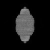 44955MC 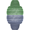 9bw5C 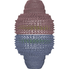 9bw7C  9mxjC M: map data used to model this data C: citing same article ( |
|---|---|
| Similar structure data |

-Links
 PDBj
PDBj


- Assembly
| Deposited unit | 
|
|---|---|
| 1 | x 39
|
| 2 |
|
| Symmetry | Point symmetry: (Schoenflies symbol: C39 (39 fold cyclic)) |
-Components
| #1: Protein | Mass: 99452.766 Da / Num. of mol.: 2 Source method: isolated from a genetically manipulated source Source: (gene. exp.) Homo sapiens (human) / Gene: MVP, LRP / Production host:  Trichoplusia ni (cabbage looper) / References: UniProt: Q14764 Trichoplusia ni (cabbage looper) / References: UniProt: Q14764#2: Protein | Mass: 192810.172 Da / Num. of mol.: 2 Source method: isolated from a genetically manipulated source Source: (gene. exp.) Homo sapiens (human) / Gene: PARP4, ADPRTL1, KIAA0177, PARPL / Production host: Trichoplusia ni (cabbage looper)References: UniProt: Q9UKK3, Transferases; Glycosyltransferases; Pentosyltransferases Has protein modification | N | |
|---|
-Experimental details
-Experiment
| Experiment | Method: ELECTRON MICROSCOPY |
|---|---|
| EM experiment | Aggregation state: PARTICLE / 3D reconstruction method: single particle reconstruction |
- Sample preparation
| Component | Name: Binary complex of human PARP4 bound to oligomerized human MVP Type: COMPLEX / Entity ID: all / Source: RECOMBINANT | |||||||||||||||||||||||||
|---|---|---|---|---|---|---|---|---|---|---|---|---|---|---|---|---|---|---|---|---|---|---|---|---|---|---|
| Molecular weight | Value: 12.7 MDa / Experimental value: NO | |||||||||||||||||||||||||
| Source (natural) | Organism: Homo sapiens (human) | |||||||||||||||||||||||||
| Source (recombinant) | Organism: Trichoplusia ni (cabbage looper) | |||||||||||||||||||||||||
| Buffer solution | pH: 8 / Details: 50 mM HEPES, 5 mM MgCl2, 5 mM CaCl2, 0.25 mM DTT | |||||||||||||||||||||||||
| Buffer component |
| |||||||||||||||||||||||||
| Specimen | Conc.: 1.5 mg/ml / Embedding applied: NO / Shadowing applied: NO / Staining applied: NO / Vitrification applied: YES / Details: Sample was monodisperse | |||||||||||||||||||||||||
| Specimen support | Details: Quantifoil grids with a 2 nm continuous carbon coating were subjected to a 15 s, 5W plasma cleaning program in O2 gas Grid material: COPPER / Grid mesh size: 200 divisions/in. / Grid type: Quantifoil R1.2/1.3 | |||||||||||||||||||||||||
| Vitrification | Instrument: FEI VITROBOT MARK IV / Cryogen name: ETHANE / Humidity: 100 % / Chamber temperature: 281 K Details: Vitrification carried out under standard conditions |
- Electron microscopy imaging
| Experimental equipment |  Model: Titan Krios / Image courtesy: FEI Company |
|---|---|
| Microscopy | Model: FEI TITAN KRIOS / Details: Preliminary grid screening was performed manually. |
| Electron gun | Electron source:  FIELD EMISSION GUN / Accelerating voltage: 300 kV / Illumination mode: FLOOD BEAM FIELD EMISSION GUN / Accelerating voltage: 300 kV / Illumination mode: FLOOD BEAM |
| Electron lens | Mode: BRIGHT FIELD / Nominal magnification: 53000 X / Nominal defocus max: 1700 nm / Nominal defocus min: 700 nm / Cs: 0.01 mm |
| Specimen holder | Cryogen: NITROGEN / Specimen holder model: FEI TITAN KRIOS AUTOGRID HOLDER |
| Image recording | Electron dose: 45 e/Å2 / Film or detector model: GATAN K3 BIOQUANTUM (6k x 4k) / Num. of real images: 7491 Details: Images were collected in movie mode, with 40 frames per image. Data was collected in SerialEM using a strategy of 3 by 3 + 1 shots per hole. |
| EM imaging optics | Energyfilter name: GIF Bioquantum / Energyfilter slit width: 8 eV Spherical aberration corrector: Microscope was modified with a Cs corrector with two hexapole elements |
- Processing
| EM software |
| ||||||||||||||||||||||||||||||||||||||||||||||||||||||||||||||||||||||||
|---|---|---|---|---|---|---|---|---|---|---|---|---|---|---|---|---|---|---|---|---|---|---|---|---|---|---|---|---|---|---|---|---|---|---|---|---|---|---|---|---|---|---|---|---|---|---|---|---|---|---|---|---|---|---|---|---|---|---|---|---|---|---|---|---|---|---|---|---|---|---|---|---|---|
| CTF correction | Details: CTF correction was performed when movies were imported into cryoSPARC Live Type: PHASE FLIPPING AND AMPLITUDE CORRECTION | ||||||||||||||||||||||||||||||||||||||||||||||||||||||||||||||||||||||||
| Particle selection | Num. of particles selected: 670079 | ||||||||||||||||||||||||||||||||||||||||||||||||||||||||||||||||||||||||
| Symmetry | Point symmetry: D39 (2x39 fold dihedral) | ||||||||||||||||||||||||||||||||||||||||||||||||||||||||||||||||||||||||
| 3D reconstruction | Resolution: 2.9 Å / Resolution method: FSC 0.143 CUT-OFF / Num. of particles: 44443 / Algorithm: BACK PROJECTION / Num. of class averages: 1 / Symmetry type: POINT | ||||||||||||||||||||||||||||||||||||||||||||||||||||||||||||||||||||||||
| Atomic model building |
| ||||||||||||||||||||||||||||||||||||||||||||||||||||||||||||||||||||||||
| Atomic model building | Chain-ID: A
|

