 Movie
Movie Controller


+ Open data

- Basic information
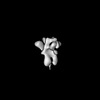
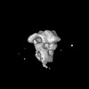
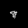
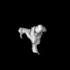
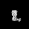
| Entry | Database: PDB / ID: 9bto | ||||||||||||||||||||||||||||||
|---|---|---|---|---|---|---|---|---|---|---|---|---|---|---|---|---|---|---|---|---|---|---|---|---|---|---|---|---|---|---|---|
| Title | Influenza hemagglutinin B/Maryland/2016 glycoprotein | ||||||||||||||||||||||||||||||
 Components Components |
| ||||||||||||||||||||||||||||||
 Keywords Keywords | VIRAL PROTEIN / hemagglutinin / influenza / complex | ||||||||||||||||||||||||||||||
| Function / homology |  Function and homology information Function and homology informationhost cell surface receptor binding / endocytosis involved in viral entry into host cell / fusion of virus membrane with host plasma membrane / fusion of virus membrane with host endosome membrane / viral envelope / virion attachment to host cell / host cell plasma membrane / virion membrane Similarity search - Function | ||||||||||||||||||||||||||||||
| Biological species |  Influenza B virus Influenza B virus | ||||||||||||||||||||||||||||||
| Method | ELECTRON MICROSCOPY / single particle reconstruction / cryo EM / Resolution: 3.1 Å | ||||||||||||||||||||||||||||||
 Authors Authors | Torrents de la Pena, A. / Sewall, L.M. / Ward, A.B. | ||||||||||||||||||||||||||||||
| Funding support |  United States, 2items United States, 2items
| ||||||||||||||||||||||||||||||
 Citation Citation | Journal: Nat Biomed Eng / Year: 2025 Title: Rapid and High-Throughput Imaging of Immune Complexes Using Microfluidics and Single Particle Electron Microscopy Authors: Sewall, L.M. / Torrents de la Pena, A. / Ward, A.B. | ||||||||||||||||||||||||||||||
| History |
|
- Structure visualization
| Structure viewer | Molecule: MolmilJmol/JSmol |
|---|
- Downloads & links
-Download
| PDBx/mmCIF format | 9bto.cif.gz | 297.2 KB | Display | PDBx/mmCIF format |
|---|---|---|---|---|
| PDB format | pdb9bto.ent.gz | 240.4 KB | Display | PDB format |
| PDBx/mmJSON format | 9bto.json.gz | Tree view | PDBx/mmJSON format | |
| Others |  Other downloads Other downloads |
-Validation report
| Arichive directory | https://data.pdbj.org/pub/pdb/validation_reports/bt/9btoftp://data.pdbj.org/pub/pdb/validation_reports/bt/9bto | HTTPS FTP |
|---|
-Related structure data
| Related structure data |  44894MC  9blkC M: map data used to model this data C: citing same article ( |
|---|---|
| Similar structure data |


-Links
 PDBj
PDBj


- Assembly
| Deposited unit | 
|
|---|---|
| 1 |
|
-Components
| #1: Protein | Mass: 39957.883 Da / Num. of mol.: 3 Source method: isolated from a genetically manipulated source Source: (gene. exp.) Influenza B virus / Strain: B/Maryland/2016 / Gene: HA / Production host:  Homo sapiens (human) / References: UniProt: A0A2Z5DTY0 Homo sapiens (human) / References: UniProt: A0A2Z5DTY0#2: Protein | Mass: 24825.535 Da / Num. of mol.: 3 Source method: isolated from a genetically manipulated source Source: (gene. exp.) Influenza B virus / Strain: B/Maryland/2016 / Gene: HA / Production host: Homo sapiens (human) / References: UniProt: A0A2Z5DTY0#3: Polysaccharide | beta-D-mannopyranose-(1-4)-2-acetamido-2-deoxy-beta-D-glucopyranose-(1-4)-2-acetamido-2-deoxy-beta- ...beta-D-mannopyranose-(1-4)-2-acetamido-2-deoxy-beta-D-glucopyranose-(1-4)-2-acetamido-2-deoxy-beta-D-glucopyranose Source method: isolated from a genetically manipulated source #4: Polysaccharide | 2-acetamido-2-deoxy-beta-D-glucopyranose-(1-4)-2-acetamido-2-deoxy-beta-D-glucopyranose Source method: isolated from a genetically manipulated source #5: Sugar | ChemComp-NAG /   Type: D-saccharide, beta linking / Mass: 221.208 Da / Num. of mol.: 6 / Source method: obtained synthetically / Formula: C8H15NO6 Type: D-saccharide, beta linking / Mass: 221.208 Da / Num. of mol.: 6 / Source method: obtained synthetically / Formula: C8H15NO6Has ligand of interest | N | Has protein modification | Y | |
|---|
-Experimental details
-Experiment
| Experiment | Method: ELECTRON MICROSCOPY |
|---|---|
| EM experiment | Aggregation state: PARTICLE / 3D reconstruction method: single particle reconstruction |
- Sample preparation
| Component | Name: influenza hemagglutinin B/Maryland/2016 glycoprotein / Type: COMPLEX / Details: HA1 and HA2 are two subunits of the same protomer / Entity ID: #1 / Source: RECOMBINANT |
|---|---|
| Source (natural) | Organism: Influenza B virus |
| Source (recombinant) | Organism: Homo sapiens (human) |
| Buffer solution | pH: 7.4 / Details: TBS |
| Specimen | Conc.: 0.02 mg/ml / Embedding applied: NO / Shadowing applied: NO / Staining applied: NO / Vitrification applied: YES |
| Specimen support | Grid type: EMS Lacey Carbon |
| Vitrification | Instrument: FEI VITROBOT MARK IV / Cryogen name: ETHANE / Humidity: 100 % / Chamber temperature: 277 K Details: wait time 30 sec, double blot for 4.5s, blot force 1 |
- Electron microscopy imaging
| Microscopy | Model: TFS GLACIOS |
|---|---|
| Electron gun | Electron source:  FIELD EMISSION GUN / Accelerating voltage: 200 kV / Illumination mode: FLOOD BEAM FIELD EMISSION GUN / Accelerating voltage: 200 kV / Illumination mode: FLOOD BEAM |
| Electron lens | Mode: BRIGHT FIELD / Nominal magnification: 190000 X / Nominal defocus max: 1800 nm / Nominal defocus min: 800 nm / Cs: 2.7 mm |
| Image recording | Electron dose: 50 e/Å2 / Film or detector model: FEI FALCON IV (4k x 4k) / Num. of real images: 815 |
- Processing
| EM software |
| ||||||||||||||||||||||||
|---|---|---|---|---|---|---|---|---|---|---|---|---|---|---|---|---|---|---|---|---|---|---|---|---|---|
| CTF correction | Type: PHASE FLIPPING AND AMPLITUDE CORRECTION | ||||||||||||||||||||||||
| 3D reconstruction | Resolution: 3.1 Å / Resolution method: FSC 0.143 CUT-OFF / Num. of particles: 16096 / Symmetry type: POINT | ||||||||||||||||||||||||
| Atomic model building | Protocol: AB INITIO MODEL | ||||||||||||||||||||||||
| Refine LS restraints |
|
