 Movie
Movie Controller

[English] 日本語
 Yorodumi
- EMDB-44667: CoV SARS-2 spike protein complexed by mEM with monoclonal antibod... -
+ Open data

- Basic information
| Entry | 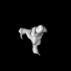 | ||||||||||||
|---|---|---|---|---|---|---|---|---|---|---|---|---|---|
| Title | CoV SARS-2 spike protein complexed by mEM with monoclonal antibody TXG-0078 Fab | ||||||||||||
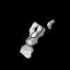
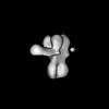
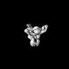
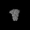
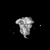
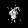
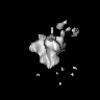
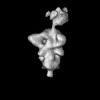
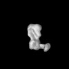
 Map data Map data | 3D refined map of SARS-2 spike with monoclonal antibody TXG-0078 Fab (C1 symmetry) | ||||||||||||
 Sample Sample |
| ||||||||||||
 Keywords Keywords | CoV / spike / SARS-2 / monoclonal antibody / TXG-0078 / complex / microfluidics / VIRAL PROTEIN | ||||||||||||
| Biological species |   Severe acute respiratory syndrome coronavirus 2 Severe acute respiratory syndrome coronavirus 2 | ||||||||||||
| Method | single particle reconstruction / negative staining / Resolution: 20.0 Å | ||||||||||||
 Authors Authors | Sewall LM / Ward AB / Torrents de la Pena A | ||||||||||||
| Funding support |  United States, 3 items United States, 3 items
| ||||||||||||
 Citation Citation | Journal: Nat Biomed Eng / Year: 2025 Title: Rapid and High-Throughput Imaging of Immune Complexes Using Microfluidics and Single Particle Electron Microscopy Authors: Sewall LM / de Paiva Froes Rocha R / Gibson G / Louie M / Xie Z / Bangaru S / Tran AS / Ozorowski G / Mohanty S / Beutler N / Burton DR / Shaw AC / Batista FD / Chocarro Ruiz B / Torrents de la Pena A / Ward AB | ||||||||||||
| History |
|
- Structure visualization
| Supplemental images |
|---|

- Downloads & links
-EMDB archive
| Map data | emd_44667.map.gz | 49.7 MB |  EMDB map data format EMDB map data format | |
|---|---|---|---|---|
| Header (meta data) | emd-44667-v30.xmlemd-44667.xml | 13.5 KB 13.5 KB | Display Display | EMDB header |
| Images |  emd_44667.png emd_44667.png | 62.1 KB | ||
| Filedesc metadata | emd-44667.cif.gz | 4 KB | ||
| Others | emd_44667_half_map_1.map.gzemd_44667_half_map_2.map.gz | 49.6 MB 49.7 MB | ||
| Archive directory |  http://ftp.pdbj.org/pub/emdb/structures/EMD-44667ftp://ftp.pdbj.org/pub/emdb/structures/EMD-44667 http://ftp.pdbj.org/pub/emdb/structures/EMD-44667ftp://ftp.pdbj.org/pub/emdb/structures/EMD-44667 | HTTPS FTP |
-Related structure data


-Links
| EMDB pages | EMDB (EBI/PDBe) / EMDataResource |
|---|
-Map
| File | Download / File: emd_44667.map.gz / Format: CCP4 / Size: 64 MB / Type: IMAGE STORED AS FLOATING POINT NUMBER (4 BYTES) | ||||||||||||||||||||||||||||||||||||
|---|---|---|---|---|---|---|---|---|---|---|---|---|---|---|---|---|---|---|---|---|---|---|---|---|---|---|---|---|---|---|---|---|---|---|---|---|---|
| Annotation | 3D refined map of SARS-2 spike with monoclonal antibody TXG-0078 Fab (C1 symmetry) | ||||||||||||||||||||||||||||||||||||
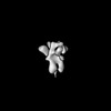
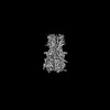
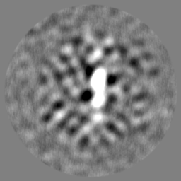
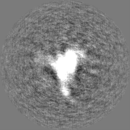
| Projections & slices | Image control
Images are generated by Spider. | ||||||||||||||||||||||||||||||||||||
| Voxel size | X=Y=Z: 2.06 Å | ||||||||||||||||||||||||||||||||||||
| Density |
| ||||||||||||||||||||||||||||||||||||
| Symmetry | Space group: 1 | ||||||||||||||||||||||||||||||||||||
| Details | EMDB XML:
|
 Z (Sec.)
 Y (Row.)
 X (Col.)


-Supplemental data
-Half map: 3D refined half map 1 of SARS-2 spike...
| File | emd_44667_half_map_1.map | ||||||||||||
|---|---|---|---|---|---|---|---|---|---|---|---|---|---|
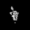
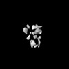
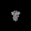
| Annotation | 3D refined half map 1 of SARS-2 spike with monoclonal antibody TXG-0078 Fab (C1 symmetry) | ||||||||||||
| Projections & Slices |
| ||||||||||||
| Density Histograms |


-Half map: 3D refined half map 2 of SARS-2 spike...
| File | emd_44667_half_map_2.map | ||||||||||||
|---|---|---|---|---|---|---|---|---|---|---|---|---|---|
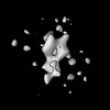
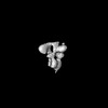
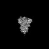
| Annotation | 3D refined half map 2 of SARS-2 spike with monoclonal antibody TXG-0078 Fab (C1 symmetry) | ||||||||||||
| Projections & Slices |
| ||||||||||||
| Density Histograms |


- Sample components
-Entire : Coronavirus SARS-2 spike in complex with monoclonal antibody TXG-...
| Entire | Name: Coronavirus SARS-2 spike in complex with monoclonal antibody TXG-0078 by mEM |
|---|---|
| Components |
|
-Supramolecule #1: Coronavirus SARS-2 spike in complex with monoclonal antibody TXG-...
| Supramolecule | Name: Coronavirus SARS-2 spike in complex with monoclonal antibody TXG-0078 by mEM type: complex / ID: 1 / Parent: 0 |
|---|---|
| Source (natural) | Organism: Severe acute respiratory syndrome coronavirus 2 |
-Experimental details
-Structure determination
| Method | negative staining |
|---|---|
 Processing Processing | single particle reconstruction |
| Aggregation state | particle |
-Sample preparation
| Buffer | pH: 7.4 / Details: 10 mM Tris, 150 mM NaCl |
|---|---|
| Staining | Type: NEGATIVE / Material: Uranyl Formate |
- Electron microscopy
| Microscope | FEI TECNAI SPIRIT |
|---|---|
| Image recording | Film or detector model: FEI EAGLE (4k x 4k) / Average electron dose: 50.0 e/Å2 |
| Electron beam | Acceleration voltage: 120 kV / Electron source:  FIELD EMISSION GUN FIELD EMISSION GUN |
| Electron optics | C2 aperture diameter: 70.0 µm / Illumination mode: FLOOD BEAM / Imaging mode: BRIGHT FIELD / Nominal defocus max: 2.0 µm / Nominal defocus min: 1.5 µm / Nominal magnification: 56000 |
| Sample stage | Specimen holder model: SIDE ENTRY, EUCENTRIC / Cooling holder cryogen: NITROGEN |
| Experimental equipment |  Model: Tecnai Spirit / Image courtesy: FEI Company |
-Image processing
| Startup model | Type of model: PDB ENTRY PDB model - PDB ID: |
|---|---|
| Final reconstruction | Resolution.type: BY AUTHOR / Resolution: 20.0 Å / Resolution method: FSC 0.143 CUT-OFF / Number images used: 4000 |
| Initial angle assignment | Type: ANGULAR RECONSTITUTION |
| Final angle assignment | Type: ANGULAR RECONSTITUTION |

