 Movie
Movie Controller


+ Open data

- Basic information
| Entry | 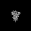 | |||||||||
|---|---|---|---|---|---|---|---|---|---|---|
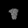
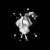
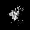
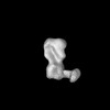
| Title | SARS-Cov-2 spike glycoprotein with 1 RBD up | |||||||||
 Map data Map data | sharpened map of SARS-2 S glycoprotein in open state | |||||||||
 Sample Sample |
| |||||||||
 Keywords Keywords | SARS-CoV-2 / open state / coronavirus / VIRAL PROTEIN | |||||||||
| Biological species |   Severe acute respiratory syndrome coronavirus 2 Severe acute respiratory syndrome coronavirus 2 | |||||||||
| Method | single particle reconstruction / cryo EM / Resolution: 6.9 Å | |||||||||
 Authors Authors | Torrents de la Pena A / Sewall LM / Ward AB | |||||||||
| Funding support |  United States, 2 items United States, 2 items
| |||||||||
 Citation Citation | Journal: Nat Biomed Eng / Year: 2025 Title: Rapid and High-Throughput Imaging of Immune Complexes Using Microfluidics and Single Particle Electron Microscopy Authors: Sewall LM / de Paiva Froes Rocha R / Gibson G / Louie M / Xie Z / Bangaru S / Tran AS / Ozorowski G / Mohanty S / Beutler N / Burton DR / Shaw AC / Batista FD / Chocarro Ruiz B / Torrents de la Pena A / Ward AB | |||||||||
| History |
|
- Structure visualization
| Supplemental images |
|---|

- Downloads & links
-EMDB archive
| Map data | emd_44680.map.gz | 229.9 MB |  EMDB map data format EMDB map data format | |
|---|---|---|---|---|
| Header (meta data) | emd-44680-v30.xmlemd-44680.xml | 15.9 KB 15.9 KB | Display Display | EMDB header |
| FSC (resolution estimation) | emd_44680_fsc.xml | 13.4 KB | Display | FSC data file |
| Images |  emd_44680.png emd_44680.png | 20 KB | ||
| Masks | emd_44680_msk_1.map | 244.1 MB | Mask map | |
| Filedesc metadata | emd-44680.cif.gz | 4.3 KB | ||
| Others | emd_44680_additional_1.map.gzemd_44680_half_map_1.map.gzemd_44680_half_map_2.map.gz | 120.2 MB 226.7 MB 226.7 MB | ||
| Archive directory |  http://ftp.pdbj.org/pub/emdb/structures/EMD-44680ftp://ftp.pdbj.org/pub/emdb/structures/EMD-44680 http://ftp.pdbj.org/pub/emdb/structures/EMD-44680ftp://ftp.pdbj.org/pub/emdb/structures/EMD-44680 | HTTPS FTP |
-Related structure data


-Links
| EMDB pages | EMDB (EBI/PDBe) / EMDataResource |
|---|
-Map
| File | Download / File: emd_44680.map.gz / Format: CCP4 / Size: 244.1 MB / Type: IMAGE STORED AS FLOATING POINT NUMBER (4 BYTES) | ||||||||||||||||||||||||||||||||||||
|---|---|---|---|---|---|---|---|---|---|---|---|---|---|---|---|---|---|---|---|---|---|---|---|---|---|---|---|---|---|---|---|---|---|---|---|---|---|
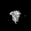
| Annotation | sharpened map of SARS-2 S glycoprotein in open state | ||||||||||||||||||||||||||||||||||||
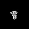
| Projections & slices | Image control
Images are generated by Spider. | ||||||||||||||||||||||||||||||||||||
| Voxel size | X=Y=Z: 1.15 Å | ||||||||||||||||||||||||||||||||||||
| Density |
| ||||||||||||||||||||||||||||||||||||
| Symmetry | Space group: 1 | ||||||||||||||||||||||||||||||||||||
| Details | EMDB XML:
|
 Z (Sec.)
 Y (Row.)
 X (Col.)


-Supplemental data
-Mask #1
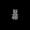
| File | emd_44680_msk_1.map | ||||||||||||
|---|---|---|---|---|---|---|---|---|---|---|---|---|---|
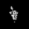
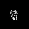
| Projections & Slices |
| ||||||||||||
| Density Histograms |


-Additional map: unsharpened map of SARS-2 S glycoprotein in open state
| File | emd_44680_additional_1.map | ||||||||||||
|---|---|---|---|---|---|---|---|---|---|---|---|---|---|
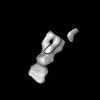
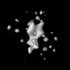
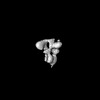
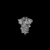
| Annotation | unsharpened map of SARS-2 S glycoprotein in open state | ||||||||||||
| Projections & Slices |
| ||||||||||||
| Density Histograms |


-Half map: half map SARS-2 S glycoprotein in open state
| File | emd_44680_half_map_1.map | ||||||||||||
|---|---|---|---|---|---|---|---|---|---|---|---|---|---|
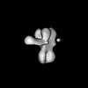
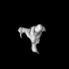
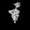
| Annotation | half map SARS-2 S glycoprotein in open state | ||||||||||||
| Projections & Slices |
| ||||||||||||
| Density Histograms |


-Half map: half map of SARS-2 S glycoprotein in open state
| File | emd_44680_half_map_2.map | ||||||||||||
|---|---|---|---|---|---|---|---|---|---|---|---|---|---|
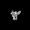
| Annotation | half map of SARS-2 S glycoprotein in open state | ||||||||||||
| Projections & Slices |
| ||||||||||||
| Density Histograms |


- Sample components
-Entire : SARS-CoV-2 S glycoprotein containing 2P mutations with 1 RBD up
| Entire | Name: SARS-CoV-2 S glycoprotein containing 2P mutations with 1 RBD up |
|---|---|
| Components |
|
-Supramolecule #1: SARS-CoV-2 S glycoprotein containing 2P mutations with 1 RBD up
| Supramolecule | Name: SARS-CoV-2 S glycoprotein containing 2P mutations with 1 RBD up type: complex / ID: 1 / Parent: 0 |
|---|---|
| Source (natural) | Organism: Severe acute respiratory syndrome coronavirus 2 |
-Experimental details
-Structure determination
| Method | cryo EM |
|---|---|
 Processing Processing | single particle reconstruction |
| Aggregation state | particle |
-Sample preparation
| Concentration | 0.02 mg/mL |
|---|---|
| Buffer | pH: 7.4 |
| Grid | Model: EMS Lacey Carbon / Material: GRAPHENE OXIDE / Mesh: 300 / Support film - Material: GRAPHENE OXIDE / Support film - topology: CONTINUOUS |
| Vitrification | Cryogen name: ETHANE / Chamber humidity: 100 % / Chamber temperature: 277 K / Instrument: FEI VITROBOT MARK IV Details: 2.5 seconds blot time, 30sec waiting time, blot force 1. |
- Electron microscopy
| Microscope | FEI TALOS ARCTICA |
|---|---|
| Image recording | Film or detector model: GATAN K2 SUMMIT (4k x 4k) / Detector mode: COUNTING / Average electron dose: 50.0 e/Å2 |
| Electron beam | Acceleration voltage: 200 kV / Electron source:  FIELD EMISSION GUN FIELD EMISSION GUN |
| Electron optics | Illumination mode: FLOOD BEAM / Imaging mode: BRIGHT FIELD / Nominal defocus max: 2.0 µm / Nominal defocus min: 1.0 µm |
| Experimental equipment |  Model: Talos Arctica / Image courtesy: FEI Company |

