 Movie
Movie Controller


+ Open data

- Basic information
| Entry | Database: PDB / ID: 9bpc | ||||||
|---|---|---|---|---|---|---|---|
| Title | Cryo-EM structure of P2X3 receptor in complex with camlipixant | ||||||
 Components Components | P2X purinoceptor | ||||||
 Keywords Keywords | MEMBRANE PROTEIN / P2X3 / camlipixant / cryo-EM / ion channel / receptor | ||||||
| Function / homology |  Function and homology information Function and homology informationextracellularly ATP-gated monoatomic cation channel activity / purinergic nucleotide receptor activity / response to ATP / postsynapse / ATP binding / plasma membrane Similarity search - Function | ||||||
| Biological species |  | ||||||
| Method | ELECTRON MICROSCOPY / single particle reconstruction / cryo EM / Resolution: 3.44 Å | ||||||
 Authors Authors | Thach, T. / Subramanian, R. | ||||||
| Funding support | 1items
| ||||||
 Citation Citation | Journal: J Biol Chem / Year: 2025 Title: Mechanistic insights into the selective targeting of P2X3 receptor by camlipixant antagonist. Authors: Trung Thach / KanagaVijayan Dhanabalan / Prajwal Prabhakarrao Nandekar / Seth Stauffer / Iring Heisler / Sarah Alvarado / Jonathan Snyder / Ramaswamy Subramanian /  Abstract: ATP-activated P2X3 receptors play a pivotal role in chronic cough, affecting more than 10% of the population. Despite the challenges posed by the highly conserved structure of P2X receptors, efforts ...ATP-activated P2X3 receptors play a pivotal role in chronic cough, affecting more than 10% of the population. Despite the challenges posed by the highly conserved structure of P2X receptors, efforts to develop selective drugs targeting P2X3 have led to the development of camlipixant, a potent, selective P2X3 antagonist. However, the mechanisms of receptor desensitization, ion permeation, and structural basis of camlipixant binding to P2X3 remain unclear. Here, we report a cryo-EM structure of camlipixant-bound P2X3, revealing a previously undiscovered selective drug-binding site in the receptor. Our findings also demonstrate that conformational changes in the upper body domain, including the turret and camlipixant-binding pocket, play a critical role: turret opening facilitates P2X3 channel closure to a radius of 0.7 Å, hindering cation transfer, whereas turret closure leads to channel opening. Structural and functional studies combined with molecular dynamics simulations provide a comprehensive understanding of camlipixant's selective inhibition of P2X3, offering a foundation for future drug development targeting this receptor. | ||||||
| History |
|
- Structure visualization
| Structure viewer | Molecule: MolmilJmol/JSmol |
|---|
- Downloads & links
-Download
| PDBx/mmCIF format | 9bpc.cif.gz | 197.4 KB | Display | PDBx/mmCIF format |
|---|---|---|---|---|
| PDB format | pdb9bpc.ent.gz | Display | PDB format | |
| PDBx/mmJSON format | 9bpc.json.gz | Tree view | PDBx/mmJSON format | |
| Others |  Other downloads Other downloads |
-Validation report
| Arichive directory | https://data.pdbj.org/pub/pdb/validation_reports/bp/9bpcftp://data.pdbj.org/pub/pdb/validation_reports/bp/9bpc | HTTPS FTP |
|---|
-Related structure data
| Related structure data |  44771MC 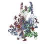 9bpdC M: map data used to model this data C: citing same article ( |
|---|---|
| Similar structure data |


-Links
 PDBj
PDBj- Assembly
| Deposited unit | 
|
|---|---|
| 1 |
|
-Components
| #1: Protein | Mass: 40477.520 Da / Num. of mol.: 3 Source method: isolated from a genetically manipulated source Source: (gene. exp.)  Homo sapiens (human) / References: UniProt: A0A8I3S575 Homo sapiens (human) / References: UniProt: A0A8I3S575#2: Polysaccharide | Source method: isolated from a genetically manipulated source #3: Chemical | Mass: 458.458 Da / Num. of mol.: 3 / Source method: obtained synthetically / Formula: C23H24F2N4O4 / Feature type: SUBJECT OF INVESTIGATION #4: Sugar |   Type: D-saccharide, beta linking / Mass: 221.208 Da / Num. of mol.: 3 / Source method: obtained synthetically / Formula: C8H15NO6 Type: D-saccharide, beta linking / Mass: 221.208 Da / Num. of mol.: 3 / Source method: obtained synthetically / Formula: C8H15NO6Has ligand of interest | Y | Has protein modification | Y | |
|---|
-Experimental details
-Experiment
| Experiment | Method: ELECTRON MICROSCOPY |
|---|---|
| EM experiment | Aggregation state: PARTICLE / 3D reconstruction method: single particle reconstruction |
- Sample preparation
| Component | Name: Ternary complex of P2X3 receptor and camlipixant / Type: COMPLEX / Details: P2X3 receptor and camlipixant antagonist / Entity ID: #1 / Source: RECOMBINANT | |||||||||||||||
|---|---|---|---|---|---|---|---|---|---|---|---|---|---|---|---|---|
| Molecular weight | Value: 0.126 MDa / Experimental value: YES | |||||||||||||||
| Source (natural) | Organism: | |||||||||||||||
| Source (recombinant) | Organism: Homo sapiens (human) | |||||||||||||||
| Buffer solution | pH: 7.5 / Details: 20 mM HEPES, 150 mM NaCl | |||||||||||||||
| Buffer component |
| |||||||||||||||
| Specimen | Conc.: 0.8 mg/ml / Embedding applied: NO / Shadowing applied: NO / Staining applied: NO / Vitrification applied: YES Details: P2X3 was incubated with camlipixant in a molar ratio of 1:5 for 30 minutes on ice before being used to prepare cryo-EM grids | |||||||||||||||
| Specimen support | Grid material: GOLD / Grid mesh size: 300 divisions/in. / Grid type: Quantifoil R1.2/1.3 | |||||||||||||||
| Vitrification | Instrument: FEI VITROBOT MARK IV / Cryogen name: ETHANE / Humidity: 100 % / Chamber temperature: 277.15 K / Details: vitrification |
- Electron microscopy imaging
| Experimental equipment |  Model: Titan Krios / Image courtesy: FEI Company |
|---|---|
| Microscopy | Model: FEI TITAN KRIOS |
| Electron gun | Electron source:  FIELD EMISSION GUN / Accelerating voltage: 300 kV / Illumination mode: FLOOD BEAM FIELD EMISSION GUN / Accelerating voltage: 300 kV / Illumination mode: FLOOD BEAM |
| Electron lens | Mode: BRIGHT FIELD / Nominal defocus max: 2000 nm / Nominal defocus min: 700 nm / Cs: 0.1 mm / C2 aperture diameter: 70 µm |
| Specimen holder | Cryogen: NITROGEN |
| Image recording | Average exposure time: 1.8 sec. / Electron dose: 56.8 e/Å2 / Film or detector model: GATAN K3 BIOCONTINUUM (6k x 4k) / Num. of grids imaged: 1 / Num. of real images: 60 |
| EM imaging optics | Energyfilter name: GIF Bioquantum |
- Processing
| EM software |
| ||||||||||||||||||||||||
|---|---|---|---|---|---|---|---|---|---|---|---|---|---|---|---|---|---|---|---|---|---|---|---|---|---|
| CTF correction | Type: PHASE FLIPPING AND AMPLITUDE CORRECTION | ||||||||||||||||||||||||
| Particle selection | Num. of particles selected: 200982 | ||||||||||||||||||||||||
| Symmetry | Point symmetry: C3 (3 fold cyclic) | ||||||||||||||||||||||||
| 3D reconstruction | Resolution: 3.44 Å / Resolution method: FSC 0.143 CUT-OFF / Num. of particles: 40142 / Num. of class averages: 50 / Symmetry type: POINT | ||||||||||||||||||||||||
| Atomic model building | Protocol: AB INITIO MODEL / Space: REAL / Details: real refinement was done using Phenix | ||||||||||||||||||||||||
| Atomic model building | Chain residue range: 1-361 / Details: The initial model consisted of monomer / Source name: AlphaFold / Type: in silico model | ||||||||||||||||||||||||
| Refine LS restraints |
|
