 Movie
Movie Controller


+ Open data

- Basic information
| Entry | 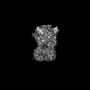 | |||||||||
|---|---|---|---|---|---|---|---|---|---|---|
| Title | Cryo-EM structure of P2X3 receptor in complex with ATP:Mg2+ | |||||||||
 Map data Map data | structure of P2X3 receptor in complex with ATP:Mg2+ | |||||||||
 Sample Sample |
| |||||||||
 Keywords Keywords | P2X3 / camlipixant / cryo-EM / ion channel / receptor / MEMBRANE PROTEIN | |||||||||
| Function / homology |  Function and homology information Function and homology informationextracellularly ATP-gated monoatomic cation channel activity / purinergic nucleotide receptor activity / response to ATP / postsynapse / ATP binding / plasma membrane Similarity search - Function | |||||||||
| Biological species |  | |||||||||
| Method | single particle reconstruction / cryo EM / Resolution: 3.63 Å | |||||||||
 Authors Authors | Thach T / Subramanian R | |||||||||
| Funding support | 1 items
| |||||||||
 Citation Citation | Journal: J Biol Chem / Year: 2025 Title: Mechanistic insights into the selective targeting of P2X3 receptor by camlipixant antagonist. Authors: Trung Thach / KanagaVijayan Dhanabalan / Prajwal Prabhakarrao Nandekar / Seth Stauffer / Iring Heisler / Sarah Alvarado / Jonathan Snyder / Ramaswamy Subramanian /  Abstract: ATP-activated P2X3 receptors play a pivotal role in chronic cough, affecting more than 10% of the population. Despite the challenges posed by the highly conserved structure of P2X receptors, efforts ...ATP-activated P2X3 receptors play a pivotal role in chronic cough, affecting more than 10% of the population. Despite the challenges posed by the highly conserved structure of P2X receptors, efforts to develop selective drugs targeting P2X3 have led to the development of camlipixant, a potent, selective P2X3 antagonist. However, the mechanisms of receptor desensitization, ion permeation, and structural basis of camlipixant binding to P2X3 remain unclear. Here, we report a cryo-EM structure of camlipixant-bound P2X3, revealing a previously undiscovered selective drug-binding site in the receptor. Our findings also demonstrate that conformational changes in the upper body domain, including the turret and camlipixant-binding pocket, play a critical role: turret opening facilitates P2X3 channel closure to a radius of 0.7 Å, hindering cation transfer, whereas turret closure leads to channel opening. Structural and functional studies combined with molecular dynamics simulations provide a comprehensive understanding of camlipixant's selective inhibition of P2X3, offering a foundation for future drug development targeting this receptor. | |||||||||
| History |
|
- Structure visualization
| Supplemental images |
|---|

- Downloads & links
-EMDB archive
| Map data | emd_44772.map.gz | 59.6 MB | EMDB map data format | |
|---|---|---|---|---|
| Header (meta data) | emd-44772-v30.xmlemd-44772.xml | 19.6 KB 19.6 KB | Display Display | EMDB header |
| FSC (resolution estimation) | emd_44772_fsc.xml | 8.5 KB | Display | FSC data file |
| Images |  emd_44772.png emd_44772.png | 45.5 KB | ||
| Filedesc metadata | emd-44772.cif.gz | 6.7 KB | ||
| Others | emd_44772_half_map_1.map.gzemd_44772_half_map_2.map.gz | 59.4 MB 59.4 MB | ||
| Archive directory |  http://ftp.pdbj.org/pub/emdb/structures/EMD-44772ftp://ftp.pdbj.org/pub/emdb/structures/EMD-44772 http://ftp.pdbj.org/pub/emdb/structures/EMD-44772ftp://ftp.pdbj.org/pub/emdb/structures/EMD-44772 | HTTPS FTP |
-Related structure data
| Related structure data | 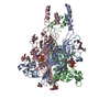 9bpdMC 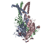 9bpcC M: atomic model generated by this map C: citing same article ( |
|---|---|
| Similar structure data |
-Links
| EMDB pages | EMDB (EBI/PDBe) / EMDataResource |
|---|
-Map
| File | Download / File: emd_44772.map.gz / Format: CCP4 / Size: 64 MB / Type: IMAGE STORED AS FLOATING POINT NUMBER (4 BYTES) | ||||||||||||||||||||||||||||||||||||
|---|---|---|---|---|---|---|---|---|---|---|---|---|---|---|---|---|---|---|---|---|---|---|---|---|---|---|---|---|---|---|---|---|---|---|---|---|---|
| Annotation | structure of P2X3 receptor in complex with ATP:Mg2+ | ||||||||||||||||||||||||||||||||||||
| Projections & slices | Image control
Images are generated by Spider. | ||||||||||||||||||||||||||||||||||||
| Voxel size | X=Y=Z: 1.054 Å | ||||||||||||||||||||||||||||||||||||
| Density |
| ||||||||||||||||||||||||||||||||||||
| Symmetry | Space group: 1 | ||||||||||||||||||||||||||||||||||||
| Details | EMDB XML:
|
 Z (Sec.)
 Y (Row.)
 X (Col.)


-Supplemental data
-Half map: Half Map A
| File | emd_44772_half_map_1.map | ||||||||||||
|---|---|---|---|---|---|---|---|---|---|---|---|---|---|
| Annotation | Half Map A | ||||||||||||
| Projections & Slices |
| ||||||||||||
| Density Histograms |


-Half map: Half Map B
| File | emd_44772_half_map_2.map | ||||||||||||
|---|---|---|---|---|---|---|---|---|---|---|---|---|---|
| Annotation | Half Map B | ||||||||||||
| Projections & Slices |
| ||||||||||||
| Density Histograms |


- Sample components
-Entire : Ternary complex of P2X3 receptor and ATP
| Entire | Name: Ternary complex of P2X3 receptor and ATP |
|---|---|
| Components |
|
-Supramolecule #1: Ternary complex of P2X3 receptor and ATP
| Supramolecule | Name: Ternary complex of P2X3 receptor and ATP / type: complex / ID: 1 / Parent: 0 / Macromolecule list: #1 / Details: P2X3 receptor and ATP |
|---|---|
| Source (natural) | Organism: |
| Molecular weight | Theoretical: 126 KDa |
-Macromolecule #1: P2X purinoceptor
| Macromolecule | Name: P2X purinoceptor / type: protein_or_peptide / ID: 1 / Number of copies: 3 / Enantiomer: LEVO |
|---|---|
| Source (natural) | Organism: |
| Molecular weight | Theoretical: 40.47752 KDa |
| Recombinant expression | Organism:  Homo sapiens (human) Homo sapiens (human) |
| Sequence | String: GSDFFTYETT KSVVVKSWTI GVINRAVQLL IISYFVGWVF LHEKAYQVRD TAIESSVVTK VKGFGRYANR VMDVSDYVTP PQGTSVFVI ITKMIVTENQ MQGFCPESEE KYRCVSDSQC GPQRFPGGGI LTGRCVNYSS TLRTCEIQGW CPTEVDTVEM P VMMEAENF ...String: GSDFFTYETT KSVVVKSWTI GVINRAVQLL IISYFVGWVF LHEKAYQVRD TAIESSVVTK VKGFGRYANR VMDVSDYVTP PQGTSVFVI ITKMIVTENQ MQGFCPESEE KYRCVSDSQC GPQRFPGGGI LTGRCVNYSS TLRTCEIQGW CPTEVDTVEM P VMMEAENF TIFIKNSIRF PLFNFEKGNL LPNLTAADMK TCRFHPDKAP FCPILRVGDV VKFAGQDFAK LARTGGVLGI KI GWVCDLD RAWDQCIPKY SFTRLDGVSE KSSVSPGYNF RFAKYYKMEN GSEYRTLLKA FGIRFDVLVY GNAGKFNIIP TII SSVAAF TSVGVGTVLC DIILLNFLKG ADQYKAKKFE EVDET UniProtKB: P2X purinoceptor |
-Macromolecule #3: 2-acetamido-2-deoxy-beta-D-glucopyranose
| Macromolecule | Name: 2-acetamido-2-deoxy-beta-D-glucopyranose / type: ligand / ID: 3 / Number of copies: 3 / Formula: NAG |
|---|---|
| Molecular weight | Theoretical: 221.208 Da |
| Chemical component information |  ChemComp-NAG: |
-Macromolecule #4: MAGNESIUM ION
| Macromolecule | Name: MAGNESIUM ION / type: ligand / ID: 4 / Number of copies: 3 / Formula: MG |
|---|---|
| Molecular weight | Theoretical: 24.305 Da |
-Macromolecule #5: ADENOSINE-5'-TRIPHOSPHATE
| Macromolecule | Name: ADENOSINE-5'-TRIPHOSPHATE / type: ligand / ID: 5 / Number of copies: 3 / Formula: ATP |
|---|---|
| Molecular weight | Theoretical: 507.181 Da |
| Chemical component information |  ChemComp-ATP: |
-Experimental details
-Structure determination
| Method | cryo EM |
|---|---|
 Processing Processing | single particle reconstruction |
| Aggregation state | particle |
-Sample preparation
| Concentration | 0.8 mg/mL | |||||||||
|---|---|---|---|---|---|---|---|---|---|---|
| Buffer | pH: 7.5 Component:
Details: 20 mM HEPES, 150 mM NaCl, 0.08%DDM | |||||||||
| Grid | Model: Quantifoil R1.2/1.3 / Material: GOLD / Mesh: 300 / Support film - Material: CARBON / Support film - topology: HOLEY / Support film - Film thickness: 11 | |||||||||
| Vitrification | Cryogen name: ETHANE / Chamber humidity: 100 % / Chamber temperature: 277.15 K / Instrument: FEI VITROBOT MARK IV / Details: vitrification. | |||||||||
| Details | P2X3:ATP |
- Electron microscopy
| Microscope | FEI TITAN KRIOS |
|---|---|
| Specialist optics | Energy filter - Name: GIF Bioquantum |
| Image recording | Film or detector model: GATAN K3 BIOCONTINUUM (6k x 4k) / Number grids imaged: 1 / Number real images: 60 / Average exposure time: 1.8 sec. / Average electron dose: 56.8 e/Å2 |
| Electron beam | Acceleration voltage: 300 kV / Electron source:  FIELD EMISSION GUN FIELD EMISSION GUN |
| Electron optics | C2 aperture diameter: 70.0 µm / Illumination mode: FLOOD BEAM / Imaging mode: BRIGHT FIELD / Cs: 0.1 mm / Nominal defocus max: 2.0 µm / Nominal defocus min: 0.7000000000000001 µm |
| Sample stage | Cooling holder cryogen: NITROGEN |
| Experimental equipment |  Model: Titan Krios / Image courtesy: FEI Company |
+Image processing

-Atomic model buiding 1
| Initial model | Chain - Chain ID: A / Chain - Residue range: 1-361 / Chain - Source name: AlphaFold / Chain - Initial model type: in silico model / Details: The initial model consisted of monomer |
|---|---|
| Details | real refinement was done using Phenix |
| Refinement | Space: REAL / Protocol: AB INITIO MODEL |
| Output model | PDB-9bpd: |
