 Movie
Movie Controller

[English] 日本語
 Yorodumi
- PDB-9bi0: Streptomyces griseus Family 2B encapsulin shell with 2-methylisob... -
+ Open data

- Basic information
| Entry | Database: PDB / ID: 9bi0 | ||||||
|---|---|---|---|---|---|---|---|
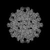
| Title | Streptomyces griseus Family 2B encapsulin shell with 2-methylisoborneol synthase cargo in 20 mM cAMP | ||||||
 Components Components | Nucleotide-binding protein | ||||||
 Keywords Keywords | VIRUS LIKE PARTICLE / Encapsulin / nanocompartment | ||||||
| Function / homology |  Function and homology information Function and homology information | ||||||
| Biological species |  Streptomyces griseus subsp. griseus (bacteria) Streptomyces griseus subsp. griseus (bacteria) | ||||||
| Method | ELECTRON MICROSCOPY / single particle reconstruction / cryo EM / Resolution: 2.93 Å | ||||||
 Authors Authors | Andreas, M.P. / Giessen, T.W. | ||||||
| Funding support |  United States, 1items United States, 1items
| ||||||
 Citation Citation | Journal: Nat Commun / Year: 2024 Title: The biosynthesis of the odorant 2-methylisoborneol is compartmentalized inside a protein shell. Authors: Michael P Andreas / Tobias W Giessen / Abstract: Terpenoids are the largest class of natural products, found across all domains of life. One of the most abundant bacterial terpenoids is the volatile odorant 2-methylisoborneol (2-MIB), partially ...Terpenoids are the largest class of natural products, found across all domains of life. One of the most abundant bacterial terpenoids is the volatile odorant 2-methylisoborneol (2-MIB), partially responsible for the earthy smell of soil and musty taste of contaminated water. Many bacterial 2-MIB biosynthetic gene clusters were thought to encode a conserved transcription factor, named EshA in the model soil bacterium Streptomyces griseus. Here, we revise the function of EshA, now referred to as Sg Enc, and show that it is a Family 2B encapsulin shell protein. Using cryo-electron microscopy, we find that Sg Enc forms an icosahedral protein shell and encapsulates 2-methylisoborneol synthase (2-MIBS) as a cargo protein. Sg Enc contains a cyclic adenosine monophosphate (cAMP) binding domain (CBD)-fold insertion and a unique metal-binding domain, both displayed on the shell exterior. We show that Sg Enc CBDs do not bind cAMP. We find that 2-MIBS cargo loading is mediated by an N-terminal disordered cargo-loading domain and that 2-MIBS activity and Sg Enc shell structure are not modulated by cAMP. Our work redefines the function of EshA and establishes Family 2B encapsulins as cargo-loaded protein nanocompartments involved in secondary metabolite biosynthesis. | ||||||
| History |
|
- Structure visualization
| Structure viewer | Molecule: MolmilJmol/JSmol |
|---|
- Downloads & links
-Download
| PDBx/mmCIF format | 9bi0.cif.gz | 85 KB | Display | PDBx/mmCIF format |
|---|---|---|---|---|
| PDB format | pdb9bi0.ent.gz | 62.3 KB | Display | PDB format |
| PDBx/mmJSON format | 9bi0.json.gz | Tree view | PDBx/mmJSON format | |
| Others |  Other downloads Other downloads |
-Validation report
| Arichive directory | https://data.pdbj.org/pub/pdb/validation_reports/bi/9bi0ftp://data.pdbj.org/pub/pdb/validation_reports/bi/9bi0 | HTTPS FTP |
|---|
-Related structure data
| Related structure data |  44557MC  9bhuC  9bhvC M: map data used to model this data C: citing same article ( |
|---|---|
| Similar structure data |

-Links
 PDBj
PDBj


- Assembly
| Deposited unit | 
|
|---|---|
| 1 | x 60
|
| 2 |
|
| 3 | x 5
|
| 4 | x 6
|
| 5 | 
|
| Symmetry | Point symmetry: (Schoenflies symbol: I (icosahedral)) |
-Components
| #1: Protein | Mass: 52014.137 Da / Num. of mol.: 1 Source method: isolated from a genetically manipulated source Details: Residues 1-39, 147-152, 216-245 are unresolved in the map. Source: (gene. exp.) Streptomyces griseus subsp. griseus (bacteria)Gene: mmpI_1, NCTC13033_02226 / Production host: Streptomyces coelicolor A3(2) (bacteria) / References: UniProt: Q54255 |
|---|---|
| #2: Chemical | ChemComp-CA /   Mass: 40.078 Da / Num. of mol.: 1 / Source method: obtained synthetically / Formula: Ca Mass: 40.078 Da / Num. of mol.: 1 / Source method: obtained synthetically / Formula: Ca |
| Has protein modification | N |
-Experimental details
-Experiment
| Experiment | Method: ELECTRON MICROSCOPY |
|---|---|
| EM experiment | Aggregation state: PARTICLE / 3D reconstruction method: single particle reconstruction |
- Sample preparation
| Component |
| ||||||||||||||||||||
|---|---|---|---|---|---|---|---|---|---|---|---|---|---|---|---|---|---|---|---|---|---|
| Molecular weight |
| ||||||||||||||||||||
| Source (natural) |
| ||||||||||||||||||||
| Source (recombinant) |
| ||||||||||||||||||||
| Buffer solution | pH: 7.5 / Details: 20 mM cAMP, 5 mM MgCl2, 20 mM HEPES pH 7.5 | ||||||||||||||||||||
| Buffer component |
| ||||||||||||||||||||
| Specimen | Conc.: 3.5 mg/ml / Embedding applied: NO / Shadowing applied: NO / Staining applied: NO / Vitrification applied: YES | ||||||||||||||||||||
| Specimen support | Details: The grid was glow discharged for 60 seconds at 5 mA under vacuum. Grid material: COPPER / Grid mesh size: 200 divisions/in. / Grid type: Quantifoil R1.2/1.3 | ||||||||||||||||||||
| Vitrification | Instrument: FEI VITROBOT MARK IV / Cryogen name: ETHANE / Humidity: 100 % / Chamber temperature: 295 K / Details: Blot force: 20 Blot time: 4 seconds |
- Electron microscopy imaging
| Experimental equipment |  Model: Talos Arctica / Image courtesy: FEI Company |
|---|---|
| Microscopy | Model: FEI TALOS ARCTICA |
| Electron gun | Electron source:  FIELD EMISSION GUN / Accelerating voltage: 200 kV / Illumination mode: FLOOD BEAM FIELD EMISSION GUN / Accelerating voltage: 200 kV / Illumination mode: FLOOD BEAM |
| Electron lens | Mode: BRIGHT FIELD / Nominal magnification: 45000 X / Nominal defocus max: 1800 nm / Nominal defocus min: 800 nm |
| Image recording | Average exposure time: 4 sec. / Electron dose: 39.98 e/Å2 / Detector mode: COUNTING / Film or detector model: GATAN K2 SUMMIT (4k x 4k) / Num. of grids imaged: 1 |
| Image scans | Width: 3710 / Height: 3838 / Movie frames/image: 20 |
- Processing
| EM software |
| ||||||||||||||||||||||||||||||||||||||||||||||||||
|---|---|---|---|---|---|---|---|---|---|---|---|---|---|---|---|---|---|---|---|---|---|---|---|---|---|---|---|---|---|---|---|---|---|---|---|---|---|---|---|---|---|---|---|---|---|---|---|---|---|---|---|
| CTF correction | Type: PHASE FLIPPING AND AMPLITUDE CORRECTION | ||||||||||||||||||||||||||||||||||||||||||||||||||
| Particle selection | Num. of particles selected: 9265 | ||||||||||||||||||||||||||||||||||||||||||||||||||
| Symmetry | Point symmetry: I (icosahedral) | ||||||||||||||||||||||||||||||||||||||||||||||||||
| 3D reconstruction | Resolution: 2.93 Å / Resolution method: FSC 0.143 CUT-OFF / Num. of particles: 9170 Details: Homogeneous refinement was performed with I symmetry, per-particle defocus optimization, per-group CTF parameterization, spherical aberration fit, tetrafoil fit, anisotropic magnification ...Details: Homogeneous refinement was performed with I symmetry, per-particle defocus optimization, per-group CTF parameterization, spherical aberration fit, tetrafoil fit, anisotropic magnification fit, and Ewald sphere correction with a positive curvature sign. Symmetry type: POINT | ||||||||||||||||||||||||||||||||||||||||||||||||||
| Atomic model building | B value: 92 / Protocol: FLEXIBLE FIT / Space: REAL / Target criteria: cross-correlation coefficient Details: An AlphaFold-generated model was fit into the map using ChimeraX v1.5. The model was then refined iteratively by alternating rounds of manual refinement in Coot v8.9.1 and real-space ...Details: An AlphaFold-generated model was fit into the map using ChimeraX v1.5. The model was then refined iteratively by alternating rounds of manual refinement in Coot v8.9.1 and real-space refinements using PHENIX v1.20.1-4487. | ||||||||||||||||||||||||||||||||||||||||||||||||||
| Atomic model building | Accession code: Q54255 / Source name: AlphaFold / Type: in silico model |
