 Movie
Movie Controller


+ Open data

- Basic information
| Entry |  | |||||||||
|---|---|---|---|---|---|---|---|---|---|---|
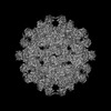
| Title | Streptomyces griseus Family 2B encapsulin shell | |||||||||
 Map data Map data | Streptomyces griseus Family 2B encapsulin shell | |||||||||
 Sample Sample |
| |||||||||
 Keywords Keywords | Encapsulin / nanocompartment / VIRUS LIKE PARTICLE | |||||||||
| Function / homology |  Function and homology information Function and homology information | |||||||||
| Biological species |  Streptomyces griseus subsp. griseus (bacteria) Streptomyces griseus subsp. griseus (bacteria) | |||||||||
| Method | single particle reconstruction / cryo EM / Resolution: 2.71 Å | |||||||||
 Authors Authors | Andreas MP / Giessen TW | |||||||||
| Funding support |  United States, 1 items United States, 1 items
| |||||||||
 Citation Citation | Journal: Nat Commun / Year: 2024 Title: The biosynthesis of the odorant 2-methylisoborneol is compartmentalized inside a protein shell. Authors: Michael P Andreas / Tobias W Giessen / Abstract: Terpenoids are the largest class of natural products, found across all domains of life. One of the most abundant bacterial terpenoids is the volatile odorant 2-methylisoborneol (2-MIB), partially ...Terpenoids are the largest class of natural products, found across all domains of life. One of the most abundant bacterial terpenoids is the volatile odorant 2-methylisoborneol (2-MIB), partially responsible for the earthy smell of soil and musty taste of contaminated water. Many bacterial 2-MIB biosynthetic gene clusters were thought to encode a conserved transcription factor, named EshA in the model soil bacterium Streptomyces griseus. Here, we revise the function of EshA, now referred to as Sg Enc, and show that it is a Family 2B encapsulin shell protein. Using cryo-electron microscopy, we find that Sg Enc forms an icosahedral protein shell and encapsulates 2-methylisoborneol synthase (2-MIBS) as a cargo protein. Sg Enc contains a cyclic adenosine monophosphate (cAMP) binding domain (CBD)-fold insertion and a unique metal-binding domain, both displayed on the shell exterior. We show that Sg Enc CBDs do not bind cAMP. We find that 2-MIBS cargo loading is mediated by an N-terminal disordered cargo-loading domain and that 2-MIBS activity and Sg Enc shell structure are not modulated by cAMP. Our work redefines the function of EshA and establishes Family 2B encapsulins as cargo-loaded protein nanocompartments involved in secondary metabolite biosynthesis. | |||||||||
| History |
|
- Structure visualization
| Supplemental images |
|---|

- Downloads & links
-EMDB archive
| Map data | emd_44553.map.gz | 306.8 MB | EMDB map data format | |
|---|---|---|---|---|
| Header (meta data) | emd-44553-v30.xmlemd-44553.xml | 20.6 KB 20.6 KB | Display Display | EMDB header |
| FSC (resolution estimation) | emd_44553_fsc.xml | 14.5 KB | Display | FSC data file |
| Images |  emd_44553.png emd_44553.png | 158.9 KB | ||
| Filedesc metadata | emd-44553.cif.gz | 6.8 KB | ||
| Others | emd_44553_additional_1.map.gzemd_44553_half_map_1.map.gzemd_44553_half_map_2.map.gz | 307.2 MB 301.4 MB 301.4 MB | ||
| Archive directory |  http://ftp.pdbj.org/pub/emdb/structures/EMD-44553ftp://ftp.pdbj.org/pub/emdb/structures/EMD-44553 http://ftp.pdbj.org/pub/emdb/structures/EMD-44553ftp://ftp.pdbj.org/pub/emdb/structures/EMD-44553 | HTTPS FTP |
-Related structure data
| Related structure data |  9bhuMC  9bhvC  9bi0C M: atomic model generated by this map C: citing same article ( |
|---|---|
| Similar structure data |

-Links
| EMDB pages | EMDB (EBI/PDBe) / EMDataResource |
|---|---|
| Related items in Molecule of the Month |


-Map
| File | Download / File: emd_44553.map.gz / Format: CCP4 / Size: 325 MB / Type: IMAGE STORED AS FLOATING POINT NUMBER (4 BYTES) | ||||||||||||||||||||||||||||||||||||
|---|---|---|---|---|---|---|---|---|---|---|---|---|---|---|---|---|---|---|---|---|---|---|---|---|---|---|---|---|---|---|---|---|---|---|---|---|---|
| Annotation | Streptomyces griseus Family 2B encapsulin shell | ||||||||||||||||||||||||||||||||||||
| Projections & slices | Image control
Images are generated by Spider. | ||||||||||||||||||||||||||||||||||||
| Voxel size | X=Y=Z: 0.91 Å | ||||||||||||||||||||||||||||||||||||
| Density |
| ||||||||||||||||||||||||||||||||||||
| Symmetry | Space group: 1 | ||||||||||||||||||||||||||||||||||||
| Details | EMDB XML:
|
 Z (Sec.)
 Y (Row.)
 X (Col.)


-Supplemental data
-Additional map: Homogeneous reconstruction (C1 symmetry) of a 3D class...
| File | emd_44553_additional_1.map | ||||||||||||
|---|---|---|---|---|---|---|---|---|---|---|---|---|---|
| Annotation | Homogeneous reconstruction (C1 symmetry) of a 3D class from focused 3D classification containing two cyclic nucleotide binding domains at the 2-fold symmetry axis in the focused mask. | ||||||||||||
| Projections & Slices |
| ||||||||||||
| Density Histograms |


-Half map: Half Map A
| File | emd_44553_half_map_1.map | ||||||||||||
|---|---|---|---|---|---|---|---|---|---|---|---|---|---|
| Annotation | Half Map A | ||||||||||||
| Projections & Slices |
| ||||||||||||
| Density Histograms |


-Half map: Half Map B
| File | emd_44553_half_map_2.map | ||||||||||||
|---|---|---|---|---|---|---|---|---|---|---|---|---|---|
| Annotation | Half Map B | ||||||||||||
| Projections & Slices |
| ||||||||||||
| Density Histograms |


- Sample components
-Entire : Streptomyces griseus Family 2B encapsulin shell
| Entire | Name: Streptomyces griseus Family 2B encapsulin shell |
|---|---|
| Components |
|
-Supramolecule #1: Streptomyces griseus Family 2B encapsulin shell
| Supramolecule | Name: Streptomyces griseus Family 2B encapsulin shell / type: complex / ID: 1 / Parent: 0 / Macromolecule list: #1 |
|---|---|
| Source (natural) | Organism: Streptomyces griseus subsp. griseus (bacteria) |
-Macromolecule #1: Nucleotide-binding protein
| Macromolecule | Name: Nucleotide-binding protein / type: protein_or_peptide / ID: 1 Details: Residues 1-39, 147-152, and 216-245 are unresolved in the map. Number of copies: 1 / Enantiomer: LEVO |
|---|---|
| Source (natural) | Organism: Streptomyces griseus subsp. griseus (bacteria) |
| Molecular weight | Theoretical: 52.014137 KDa |
| Recombinant expression | Organism: |
| Sequence | String: MTVDSTSEAR LEVPRQSSLG TAAARNLAST TKSAPQMQEI TSRWLLRMLP WVETKGGAYR VNRRLTFTVG DGRVEFVQDG STVRVIPQE LGELALLRDF DDAEVLSAIA DRCVQRDFRA GETLVERGTA ADELHLIAHG RIGQASAGSY GDEVTLDVLA D GDRFGEHA ...String: MTVDSTSEAR LEVPRQSSLG TAAARNLAST TKSAPQMQEI TSRWLLRMLP WVETKGGAYR VNRRLTFTVG DGRVEFVQDG STVRVIPQE LGELALLRDF DDAEVLSAIA DRCVQRDFRA GETLVERGTA ADELHLIAHG RIGQASAGSY GDEVTLDVLA D GDRFGEHA LLDEDARWSQ TATAETSGTL LTLSRADFAA VVANSPALRS HLAAFTARSE QRQNHRGEAE IAMSAGHVGE HE LPGAFAD YELKPREYEL SVAQTILRVH TRVADLYNGP MNQTEEQLRL TIEALRERQE HELINNREFG LLHNADFKQR IQT HSGPPT PDDLDELLCR RRGTKFFLAH PRTIAAMGRE FNARGLYPDH TDLGGQQVPA WRGVPILPCG KIPITPERTS SILA LRTGE EDQGVIGLRQ TGLPDEYEPG LSVRFMNIDE KAIISYLVST YYSAAILVPD AVGVLENVQI ANWPR UniProtKB: Sporulation protein |
-Macromolecule #2: CALCIUM ION
| Macromolecule | Name: CALCIUM ION / type: ligand / ID: 2 / Number of copies: 1 / Formula: CA |
|---|---|
| Molecular weight | Theoretical: 40.078 Da |
-Experimental details
-Structure determination
| Method | cryo EM |
|---|---|
 Processing Processing | single particle reconstruction |
| Aggregation state | particle |
-Sample preparation
| Concentration | 2.5 mg/mL | |||||||||
|---|---|---|---|---|---|---|---|---|---|---|
| Buffer | pH: 7.5 Component:
Details: 150 mM NaCl, 20 mM Tris pH 7.5 | |||||||||
| Grid | Model: Quantifoil R1.2/1.3 / Material: COPPER / Mesh: 200 / Support film - Material: CARBON / Support film - topology: HOLEY / Pretreatment - Type: GLOW DISCHARGE / Pretreatment - Time: 60 sec. / Details: 60 seconds at 5 mA under vacuum | |||||||||
| Vitrification | Cryogen name: ETHANE / Chamber humidity: 100 % / Chamber temperature: 295 K / Instrument: FEI VITROBOT MARK IV / Details: Blot force: 20 Blot time: 4 seconds. |
- Electron microscopy
| Microscope | TFS GLACIOS |
|---|---|
| Image recording | Film or detector model: GATAN K2 SUMMIT (4k x 4k) / Detector mode: COUNTING / Digitization - Dimensions - Width: 3710 pixel / Digitization - Dimensions - Height: 3838 pixel / Number grids imaged: 1 / Average exposure time: 5.0 sec. / Average electron dose: 43.22 e/Å2 |
| Electron beam | Acceleration voltage: 200 kV / Electron source:  FIELD EMISSION GUN FIELD EMISSION GUN |
| Electron optics | Illumination mode: FLOOD BEAM / Imaging mode: BRIGHT FIELD / Nominal defocus max: 1.8 µm / Nominal defocus min: 1.0 µm / Nominal magnification: 45000 |
+Image processing

-Atomic model buiding 1
| Initial model | PDB ID: Chain - Source name: AlphaFold / Chain - Initial model type: in silico model |
|---|---|
| Details | An AlphaFold-generated model was fit into the map using ChimeraX v1.5. The model was then refined iteratively by alternating rounds of manual refinement in Coot v8.9.1 and real-space refinements using PHENIX v1.20.1-4487. |
| Refinement | Space: REAL / Protocol: FLEXIBLE FIT / Overall B value: 127.9 / Target criteria: cross-correlation coefficient |
| Output model | PDB-9bhu: |

