[English] 日本語
 Yorodumi
- PDB-9bei: Cryo-EM structure of synthetic claudin-4 complex with Clostridium... -
+ Open data
- Basic information
| Entry | Database: PDB / ID: 9bei | ||||||
|---|---|---|---|---|---|---|---|
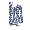
| Title | Cryo-EM structure of synthetic claudin-4 complex with Clostridium perfringens enterotoxin C-terminal domain, sFab COP-2, and Nanobody | ||||||
 Components Components |
| ||||||
 Keywords Keywords | MEMBRANE PROTEIN/IMMUNE SYSYTEM / Claudin / Fab / Toxin / MEMBRANE PROTEIN / MEMBRANE PROTEIN-IMMUNE SYSYTEM complex | ||||||
| Function / homology | Clostridium enterotoxin / Clostridium enterotoxin / toxin activity / extracellular region / Heat-labile enterotoxin B chain Function and homology information Function and homology information | ||||||
| Biological species |   Homo sapiens (human) Homo sapiens (human) | ||||||
| Method | ELECTRON MICROSCOPY / single particle reconstruction / cryo EM / Resolution: 4.16 Å | ||||||
 Authors Authors | Vecchio, A.J. | ||||||
| Funding support |  United States, 1items United States, 1items
| ||||||
 Citation Citation |  Journal: Nature / Year: 2024 Journal: Nature / Year: 2024Title: Computational design of soluble and functional membrane protein analogues. Authors: Casper A Goverde / Martin Pacesa / Nicolas Goldbach / Lars J Dornfeld / Petra E M Balbi / Sandrine Georgeon / Stéphane Rosset / Srajan Kapoor / Jagrity Choudhury / Justas Dauparas / ...Authors: Casper A Goverde / Martin Pacesa / Nicolas Goldbach / Lars J Dornfeld / Petra E M Balbi / Sandrine Georgeon / Stéphane Rosset / Srajan Kapoor / Jagrity Choudhury / Justas Dauparas / Christian Schellhaas / Simon Kozlov / David Baker / Sergey Ovchinnikov / Alex J Vecchio / Bruno E Correia /   Abstract: De novo design of complex protein folds using solely computational means remains a substantial challenge. Here we use a robust deep learning pipeline to design complex folds and soluble analogues of ...De novo design of complex protein folds using solely computational means remains a substantial challenge. Here we use a robust deep learning pipeline to design complex folds and soluble analogues of integral membrane proteins. Unique membrane topologies, such as those from G-protein-coupled receptors, are not found in the soluble proteome, and we demonstrate that their structural features can be recapitulated in solution. Biophysical analyses demonstrate the high thermal stability of the designs, and experimental structures show remarkable design accuracy. The soluble analogues were functionalized with native structural motifs, as a proof of concept for bringing membrane protein functions to the soluble proteome, potentially enabling new approaches in drug discovery. In summary, we have designed complex protein topologies and enriched them with functionalities from membrane proteins, with high experimental success rates, leading to a de facto expansion of the functional soluble fold space. | ||||||
| History |
|
- Structure visualization
| Structure viewer | Molecule:  Molmil Molmil Jmol/JSmol Jmol/JSmol |
|---|
- Downloads & links
- Download
| PDBx/mmCIF format |  9bei.cif.gz 9bei.cif.gz | 161.1 KB | Display |  PDBx/mmCIF format PDBx/mmCIF format |
|---|---|---|---|---|
| PDB format |  pdb9bei.ent.gz pdb9bei.ent.gz | Display |  PDB format PDB format | |
| PDBx/mmJSON format |  9bei.json.gz 9bei.json.gz | Tree view |  PDBx/mmJSON format PDBx/mmJSON format | |
| Others |  Other downloads Other downloads |
-Validation report
| Summary document |  9bei_validation.pdf.gz 9bei_validation.pdf.gz | 1.4 MB | Display |  wwPDB validaton report wwPDB validaton report |
|---|---|---|---|---|
| Full document |  9bei_full_validation.pdf.gz 9bei_full_validation.pdf.gz | 1.4 MB | Display | |
| Data in XML |  9bei_validation.xml.gz 9bei_validation.xml.gz | 46.3 KB | Display | |
| Data in CIF |  9bei_validation.cif.gz 9bei_validation.cif.gz | 67.3 KB | Display | |
| Arichive directory |  https://data.pdbj.org/pub/pdb/validation_reports/be/9bei https://data.pdbj.org/pub/pdb/validation_reports/be/9bei ftp://data.pdbj.org/pub/pdb/validation_reports/be/9bei ftp://data.pdbj.org/pub/pdb/validation_reports/be/9bei | HTTPS FTP |
-Related structure data
| Related structure data |  44479MC  8oysC  8oyvC  8oywC  8oyxC  8oyyC M: map data used to model this data C: citing same article ( |
|---|---|
| Similar structure data | Similarity search - Function & homology  F&H Search F&H Search |
- Links
Links
- Assembly
| Deposited unit | 
|
|---|---|
| 1 |
|
- Components
| #1: Protein | Mass: 22078.986 Da / Num. of mol.: 1 / Source method: obtained synthetically / Source: (synth.)  Homo sapiens (human) Homo sapiens (human) |
|---|---|
| #2: Protein | Mass: 14591.295 Da / Num. of mol.: 1 Source method: isolated from a genetically manipulated source Source: (gene. exp.)   Trichoplusia ni (cabbage looper) / References: UniProt: P01558 Trichoplusia ni (cabbage looper) / References: UniProt: P01558 |
| #3: Antibody | Mass: 25263.010 Da / Num. of mol.: 1 / Source method: obtained synthetically / Source: (synth.)  |
| #4: Antibody | Mass: 13175.438 Da / Num. of mol.: 1 / Source method: obtained synthetically / Source: (synth.)  |
| #5: Antibody | Mass: 23517.057 Da / Num. of mol.: 1 / Source method: obtained synthetically / Source: (synth.)  |
| Has protein modification | Y |
-Experimental details
-Experiment
| Experiment | Method: ELECTRON MICROSCOPY |
|---|---|
| EM experiment | Aggregation state: PARTICLE / 3D reconstruction method: single particle reconstruction |
- Sample preparation
| Component | Name: Synthetic human claudin-4 complex with Clostridium perfringens enterotoxin C-terminal domain, sFab COP-2, and nanobody against COP-2 Type: COMPLEX Details: Assembled complex of 5 proteins (Fab is 2 proteins) expressed from insect cells and E coli Entity ID: all / Source: RECOMBINANT | |||||||||||||||
|---|---|---|---|---|---|---|---|---|---|---|---|---|---|---|---|---|
| Molecular weight | Value: 0.103 MDa / Experimental value: NO | |||||||||||||||
| Source (natural) | Organism:  Homo sapiens (human) Homo sapiens (human) | |||||||||||||||
| Source (recombinant) | Organism:  Trichoplusia ni (cabbage looper) Trichoplusia ni (cabbage looper) | |||||||||||||||
| Buffer solution | pH: 7.4 / Details: 20 mM Hepes pH 8.0, 150 mM NaCl | |||||||||||||||
| Buffer component |
| |||||||||||||||
| Specimen | Conc.: 5 mg/ml / Embedding applied: NO / Shadowing applied: NO / Staining applied: NO / Vitrification applied: YES | |||||||||||||||
| Specimen support | Details: UltraAuFoil 1.2/1.3 grids (Quantifoil) were glow discharged for 30 s at 15 mA in a Pelco easiGlow (Ted Pella Inc) instrument Grid material: GOLD / Grid mesh size: 300 divisions/in. / Grid type: UltrAuFoil R1.2/1.3 | |||||||||||||||
| Vitrification | Instrument: LEICA EM GP / Cryogen name: ETHANE / Humidity: 100 % / Chamber temperature: 278 K Details: 3.5 microL of complex was applied onto grids and blotted for 3 s at 4 degrees C under 100 percent humidity then plunge frozen into liquid ethane cooled by liquid nitrogen. |
- Electron microscopy imaging
| Microscopy | Model: TFS GLACIOS |
|---|---|
| Electron gun | Electron source:  FIELD EMISSION GUN / Accelerating voltage: 200 kV / Illumination mode: FLOOD BEAM FIELD EMISSION GUN / Accelerating voltage: 200 kV / Illumination mode: FLOOD BEAM |
| Electron lens | Mode: BRIGHT FIELD / Nominal magnification: 120000 X / Nominal defocus max: 2000 nm / Nominal defocus min: 400 nm |
| Specimen holder | Cryogen: NITROGEN / Specimen holder model: FEI TITAN KRIOS AUTOGRID HOLDER |
| Image recording | Electron dose: 49.4 e/Å2 / Film or detector model: FEI FALCON IV (4k x 4k) / Num. of grids imaged: 1 / Num. of real images: 1159 |
- Processing
| EM software |
| |||||||||||||||||||||||||||||||||||||||||||||
|---|---|---|---|---|---|---|---|---|---|---|---|---|---|---|---|---|---|---|---|---|---|---|---|---|---|---|---|---|---|---|---|---|---|---|---|---|---|---|---|---|---|---|---|---|---|---|
| CTF correction | Type: PHASE FLIPPING AND AMPLITUDE CORRECTION | |||||||||||||||||||||||||||||||||||||||||||||
| Particle selection | Num. of particles selected: 1848208 | |||||||||||||||||||||||||||||||||||||||||||||
| Symmetry | Point symmetry: C1 (asymmetric) | |||||||||||||||||||||||||||||||||||||||||||||
| 3D reconstruction | Resolution: 4.16 Å / Resolution method: FSC 0.143 CUT-OFF / Num. of particles: 21296 / Num. of class averages: 1 / Symmetry type: POINT | |||||||||||||||||||||||||||||||||||||||||||||
| Atomic model building | B value: 322 / Protocol: FLEXIBLE FIT / Space: REAL | |||||||||||||||||||||||||||||||||||||||||||||
| Atomic model building | 3D fitting-ID: 1
| |||||||||||||||||||||||||||||||||||||||||||||
| Refine LS restraints |
|
 Movie
Movie Controller


 PDBj
PDBj

