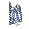
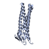
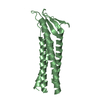
[English] 日本語
Yorodumi
- PDB-8oyv: De novo designed Claudin fold CLF_4 -

+
Open data


ID or keywords:

Loading...

-
Basic information

Entry
Database: PDB / ID: 8oyv
TitleDe novo designed Claudin fold CLF_4
ComponentsDe novo designed soluble Claudin
KeywordsDE NOVO PROTEIN / claudin / solubilized / de novo designed
Biological speciessynthetic construct (others)
MethodX-RAY DIFFRACTION / SYNCHROTRON / MOLECULAR REPLACEMENT / Resolution: 2.79 Å
AuthorsPacesa, M. / Correia, B.E.
Funding support Switzerland, European Union, 2items
OrganizationGrant numberCountry
Swiss National Science Foundation Switzerland
European Research Council (ERC)716058European Union
CitationJournal: Biorxiv / Year: 2024
Title: Computational design of soluble functional analogues of integral membrane proteins.
Authors: Goverde, C.A. / Pacesa, M. / Goldbach, N. / Dornfeld, L.J. / Balbi, P.E.M. / Georgeon, S. / Rosset, S. / Kapoor, S. / Choudhury, J. / Dauparas, J. / Schellhaas, C. / Kozlov, S. / Baker, D. / ...Authors: Goverde, C.A. / Pacesa, M. / Goldbach, N. / Dornfeld, L.J. / Balbi, P.E.M. / Georgeon, S. / Rosset, S. / Kapoor, S. / Choudhury, J. / Dauparas, J. / Schellhaas, C. / Kozlov, S. / Baker, D. / Ovchinnikov, S. / Vecchio, A.J. / Correia, B.E.
History
DepositionMay 5, 2023Deposition site: PDBE / Processing site: PDBE
Revision 1.0Oct 18, 2023Provider: repository / Type: Initial release
Revision 1.1Mar 27, 2024Group: Database references / Category: citation / citation_author
Item: _citation.country / _citation.journal_abbrev ..._citation.country / _citation.journal_abbrev / _citation.journal_id_CSD / _citation.journal_id_ISSN / _citation.pdbx_database_id_DOI / _citation.pdbx_database_id_PubMed / _citation.title / _citation.year

-
Structure visualization

Structure viewerMolecule:
MolmilJmol/JSmol

Downloads & links

-
Assembly

Deposited unit
A: De novo designed soluble Claudin
B: De novo designed soluble Claudin


Theoretical massNumber of molelcules
Total (without water)45,4212
Polymers45,4212
Non-polymers00
Water181
1
A: De novo designed soluble Claudin


Theoretical massNumber of molelcules
Total (without water)22,7111
Polymers22,7111
Non-polymers00
Water181
TypeNameSymmetry operationNumber
identity operation1_555x,y,z1
2
B: De novo designed soluble Claudin


Theoretical massNumber of molelcules
Total (without water)22,7111
Polymers22,7111
Non-polymers00
Water0
TypeNameSymmetry operationNumber
identity operation1_555x,y,z1
Unit cell
Length a, b, c (Å)35.560, 50.770, 54.930
Angle α, β, γ (deg.)107.610, 90.490, 90.060
Int Tables number1
Space group name H-MP1
Space group name HallP1
Symmetry operation#1: x,y,z
Noncrystallographic symmetry (NCS)NCS domain:
IDEns-IDDetails
d_1ens_1(chain "A" and (resid 2 through 101 or resid 108 through 186))
d_2ens_1chain "B"

NCS domain segments:
Dom-IDComponent-IDEns-IDBeg label comp-IDEnd label comp-IDLabel asym-IDLabel seq-ID
d_11ens_1GLUASNA2 - 101
d_12ens_1SERGLYA103 - 181
d_21ens_1GLUGLYB1 - 179

NCS oper: (Code: givenMatrix: (0.999966831339, 0.000295780047617, 0.00813933265649), (0.000317636741611, -0.9999963472, -0.00268415603045), (0.00813850900534, 0.00268665235169, -0.999963272611)Vector: ...NCS oper: (Code: given
Matrix: (0.999966831339, 0.000295780047617, 0.00813933265649), (0.000317636741611, -0.9999963472, -0.00268415603045), (0.00813850900534, 0.00268665235169, -0.999963272611)
Vector: -17.6814855045, -3.03858385237, 28.7760689496)

-
Components

#1: Protein De novo designed soluble Claudin


Mass: 22710.654 Da / Num. of mol.: 2
Source method: isolated from a genetically manipulated source
Source: (gene. exp.) synthetic construct (others) / Production host: Escherichia coli (E. coli)
#2: Water ChemComp-HOH / water


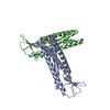
Mass: 18.015 Da / Num. of mol.: 1 / Source method: isolated from a natural source / Formula: H2O

-
Experimental details

-
Experiment

ExperimentMethod: X-RAY DIFFRACTION / Number of used crystals: 1

-
Sample preparation

CrystalDensity Matthews: 2.08 Å3/Da / Density % sol: 40.89 %
Crystal growTemperature: 277.15 K / Method: vapor diffusion, sitting drop / pH: 5
Details: 0.1 M sodium citrate pH 5, 0.1 M Sodium/Potassium phosphate pH 5.5, 0.1M Rubidium Chloride, 25% v/v PEG smear medium

-
Data collection

DiffractionMean temperature: 100 K / Serial crystal experiment: N
Diffraction sourceSource: SYNCHROTRON / Site: SLS / Beamline: X06SA / Wavelength: 1 Å
DetectorType: DECTRIS EIGER X 16M / Detector: PIXEL / Date: Apr 26, 2023
RadiationProtocol: SINGLE WAVELENGTH / Monochromatic (M) / Laue (L): M / Scattering type: x-ray
Radiation wavelengthWavelength: 1 Å / Relative weight: 1
ReflectionResolution: 2.79→52.35 Å / Num. obs: 8007 / % possible obs: 88.03 % / Redundancy: 2.5 % / Biso Wilson estimate: 77.62 Å2 / CC1/2: 0.99 / Rmerge(I) obs: 0.1069 / Net I/σ(I): 6.34
Reflection shellResolution: 2.795→2.895 Å / Redundancy: 2.7 % / Rmerge(I) obs: 1.588 / Mean I/σ(I) obs: 1.42 / Num. unique obs: 858 / CC1/2: 0.437 / % possible all: 95.12

-
Processing

Software
NameVersionClassification
PHENIX1.20.1_4487refinement
XDSdata reduction
XSCALEdata scaling
PHASERphasing
RefinementMethod to determine structure: MOLECULAR REPLACEMENT / Resolution: 2.79→52.35 Å / SU ML: 0.458 / Cross valid method: FREE R-VALUE / σ(F): 1.96 / Phase error: 42.9096
Stereochemistry target values: GeoStd + Monomer Library + CDL v1.2
RfactorNum. reflection% reflection
Rfree0.3176 367 4.6 %
Rwork0.2725 7614 -
obs0.2744 7981 88 %
Solvent computationShrinkage radii: 0.9 Å / VDW probe radii: 1.1 Å / Solvent model: FLAT BULK SOLVENT MODEL
Displacement parametersBiso mean: 97.64 Å2
Refinement stepCycle: LAST / Resolution: 2.79→52.35 Å
ProteinNucleic acidLigandSolventTotal
Num. atoms2950 0 0 1 2951
Refine LS restraints
Refine-IDTypeDev idealNumber
X-RAY DIFFRACTIONf_bond_d0.00212978
X-RAY DIFFRACTIONf_angle_d0.47713992
X-RAY DIFFRACTIONf_chiral_restr0.0364460
X-RAY DIFFRACTIONf_plane_restr0.0036508
X-RAY DIFFRACTIONf_dihedral_angle_d13.33211206
Refine LS restraints NCSType: Torsion NCS / Rms dev position: 0.423568396936 Å
LS refinement shell
Resolution (Å)Rfactor RfreeNum. reflection RfreeRfactor RworkNum. reflection RworkRefine-ID% reflection obs (%)
2.79-3.20.44281530.39662738X-RAY DIFFRACTION95.76
3.2-4.030.4034950.3292158X-RAY DIFFRACTION74.78
4.03-52.350.26111190.2312718X-RAY DIFFRACTION93.45

+
About Yorodumi

-
News

-
Feb 9, 2022. New format data for meta-information of EMDB entries

New format data for meta-information of EMDB entries

  • Version 3 of the EMDB header file is now the official format.
  • The previous official version 1.9 will be removed from the archive.

Related info.:EMDB header

External links:wwPDB to switch to version 3 of the EMDB data model

-
Aug 12, 2020. Covid-19 info

Covid-19 info

URL: https://pdbjlvh1.pdbj.org/emnavi/covid19.php

New page: Covid-19 featured information page in EM Navigator.

Related info.:Covid-19 info / Mar 5, 2020. Novel coronavirus structure data

+
Mar 5, 2020. Novel coronavirus structure data

Novel coronavirus structure data

Related info.:Yorodumi Speices / Aug 12, 2020. Covid-19 info

External links:COVID-19 featured content - PDBj / Molecule of the Month (242):Coronavirus Proteases

+
Jan 31, 2019. EMDB accession codes are about to change! (news from PDBe EMDB page)

EMDB accession codes are about to change! (news from PDBe EMDB page)

  • The allocation of 4 digits for EMDB accession codes will soon come to an end. Whilst these codes will remain in use, new EMDB accession codes will include an additional digit and will expand incrementally as the available range of codes is exhausted. The current 4-digit format prefixed with “EMD-” (i.e. EMD-XXXX) will advance to a 5-digit format (i.e. EMD-XXXXX), and so on. It is currently estimated that the 4-digit codes will be depleted around Spring 2019, at which point the 5-digit format will come into force.
  • The EM Navigator/Yorodumi systems omit the EMD- prefix.

Related info.:Q: What is EMD? / ID/Accession-code notation in Yorodumi/EM Navigator

External links:EMDB Accession Codes are Changing Soon! / Contact to PDBj

+
Jul 12, 2017. Major update of PDB

Major update of PDB

  • wwPDB released updated PDB data conforming to the new PDBx/mmCIF dictionary.
  • This is a major update changing the version number from 4 to 5, and with Remediation, in which all the entries are updated.
  • In this update, many items about electron microscopy experimental information are reorganized (e.g. em_software).
  • Now, EM Navigator and Yorodumi are based on the updated data.

External links:wwPDB Remediation / Enriched Model Files Conforming to OneDep Data Standards Now Available in the PDB FTP Archive

-
Yorodumi

Thousand views of thousand structures

  • Yorodumi is a browser for structure data from EMDB, PDB, SASBDB, etc.
  • This page is also the successor to EM Navigator detail page, and also detail information page/front-end page for Omokage search.
  • The word "yorodu" (or yorozu) is an old Japanese word meaning "ten thousand". "mi" (miru) is to see.

Related info.:EMDB / PDB / SASBDB / Comparison of 3 databanks / Yorodumi Search / Aug 31, 2016. New EM Navigator & Yorodumi / Yorodumi Papers / Jmol/JSmol / Function and homology information / Changes in new EM Navigator and Yorodumi

Read more