 Movie
Movie Controller

[English] 日本語
 Yorodumi
- PDB-9b8h: Cryo-EM structure of E. coli cellulose synthase subunit C with ce... -
+ Open data

- Basic information
| Entry | Database: PDB / ID: 9b8h | ||||||||||||||||||||||||
|---|---|---|---|---|---|---|---|---|---|---|---|---|---|---|---|---|---|---|---|---|---|---|---|---|---|
| Title | Cryo-EM structure of E. coli cellulose synthase subunit C with cellotetraose | ||||||||||||||||||||||||
 Components Components | Cellulose synthase operon protein C | ||||||||||||||||||||||||
 Keywords Keywords | MEMBRANE PROTEIN / Porin / BcsC outer membrane protein / Tetra-tricopeptide (TPR) repeats / cellooligosaccharide | ||||||||||||||||||||||||
| Function / homology |  Function and homology information Function and homology informationBiofilm formation / cellulose biosynthetic process / cell outer membrane Similarity search - Function | ||||||||||||||||||||||||
| Biological species |  | ||||||||||||||||||||||||
| Method | ELECTRON MICROSCOPY / single particle reconstruction / cryo EM / Resolution: 3.8 Å | ||||||||||||||||||||||||
 Authors Authors | Verma, P. / Zimmer, J. | ||||||||||||||||||||||||
| Funding support |  United States, 1items United States, 1items
| ||||||||||||||||||||||||
 Citation Citation | Journal: Nat Commun / Year: 2024 Title: Insights into phosphoethanolamine cellulose synthesis and secretion across the Gram-negative cell envelope. Authors: Preeti Verma / Ruoya Ho / Schuyler A Chambers / Lynette Cegelski / Jochen Zimmer / Abstract: Phosphoethanolamine (pEtN) cellulose is a naturally occurring modified cellulose produced by several Enterobacteriaceae. The minimal components of the E. coli cellulose synthase complex include the ...Phosphoethanolamine (pEtN) cellulose is a naturally occurring modified cellulose produced by several Enterobacteriaceae. The minimal components of the E. coli cellulose synthase complex include the catalytically active BcsA enzyme, a hexameric semicircle of the periplasmic BcsB protein, and the outer membrane (OM)-integrated BcsC subunit containing periplasmic tetratricopeptide repeats (TPR). Additional subunits include BcsG, a membrane-anchored periplasmic pEtN transferase associated with BcsA, and BcsZ, a periplasmic cellulase of unknown biological function. While cellulose synthesis and translocation by BcsA are well described, little is known about its pEtN modification and translocation across the cell envelope. We show that the N-terminal cytosolic domain of BcsA positions three BcsG copies near the nascent cellulose polymer. Further, the semicircle's terminal BcsB subunit tethers the N-terminus of a single BcsC protein in a trans-envelope secretion system. BcsC's TPR motifs bind a putative cello-oligosaccharide near the entrance to its OM pore. Additionally, we show that only the hydrolytic activity of BcsZ but not the subunit itself is necessary for cellulose secretion, suggesting a secretion mechanism based on enzymatic removal of translocation incompetent cellulose. Lastly, protein engineering introduces cellulose pEtN modification in orthogonal cellulose biosynthetic systems. These findings advance our understanding of pEtN cellulose modification and secretion. | ||||||||||||||||||||||||
| History |
|
- Structure visualization
| Structure viewer | Molecule: MolmilJmol/JSmol |
|---|
- Downloads & links
-Download
| PDBx/mmCIF format | 9b8h.cif.gz | 128.4 KB | Display | PDBx/mmCIF format |
|---|---|---|---|---|
| PDB format | pdb9b8h.ent.gz | Display | PDB format | |
| PDBx/mmJSON format | 9b8h.json.gz | Tree view | PDBx/mmJSON format | |
| Others |  Other downloads Other downloads |
-Validation report
| Arichive directory | https://data.pdbj.org/pub/pdb/validation_reports/b8/9b8hftp://data.pdbj.org/pub/pdb/validation_reports/b8/9b8h | HTTPS FTP |
|---|
-Related structure data
| Related structure data |  44345MC  9b87C 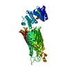 9b8aC  9b8iC  9b8vC M: map data used to model this data C: citing same article ( |
|---|---|
| Similar structure data |


-Links
 PDBj
PDBj


- Assembly
| Deposited unit | 
|
|---|---|
| 1 |
|
-Components
| #1: Protein | Mass: 126827.016 Da / Num. of mol.: 1 Source method: isolated from a genetically manipulated source Source: (gene. exp.) |
|---|---|
| #2: Polysaccharide | beta-D-glucopyranose-(1-4)-beta-D-glucopyranose-(1-4)-beta-D-glucopyranose-(1-4)-beta-D-glucopyranose |
| Has ligand of interest | Y |
| Has protein modification | Y |
-Experimental details
-Experiment
| Experiment | Method: ELECTRON MICROSCOPY |
|---|---|
| EM experiment | Aggregation state: PARTICLE / 3D reconstruction method: single particle reconstruction |
- Sample preparation
| Component | Name: Bacterial cellulose synthase subunit C in nanodisc with cellotetraose Type: COMPLEX Details: Protein: Bacterial cellulose synthase subunit C (BcsC) Ligand: Cellotetraose Entity ID: #1 / Source: RECOMBINANT |
|---|---|
| Source (natural) | Organism: |
| Source (recombinant) | Organism: |
| Buffer solution | pH: 8.5 |
| Specimen | Embedding applied: NO / Shadowing applied: NO / Staining applied: NO / Vitrification applied: YES |
| Vitrification | Cryogen name: ETHANE |
- Electron microscopy imaging
| Experimental equipment |  Model: Titan Krios / Image courtesy: FEI Company |
|---|---|
| Microscopy | Model: FEI TITAN KRIOS |
| Electron gun | Electron source:  FIELD EMISSION GUN / Accelerating voltage: 300 kV / Illumination mode: FLOOD BEAM FIELD EMISSION GUN / Accelerating voltage: 300 kV / Illumination mode: FLOOD BEAM |
| Electron lens | Mode: BRIGHT FIELD / Nominal defocus max: 2400 nm / Nominal defocus min: 1200 nm |
| Image recording | Electron dose: 50 e/Å2 / Film or detector model: GATAN K3 BIOQUANTUM (6k x 4k) |
- Processing
| EM software | Name: PHENIX / Category: model refinement |
|---|---|
| CTF correction | Type: PHASE FLIPPING AND AMPLITUDE CORRECTION |
| 3D reconstruction | Resolution: 3.8 Å / Resolution method: FSC 0.143 CUT-OFF / Num. of particles: 60119 / Symmetry type: POINT |
