 Movie
Movie Controller

[English] 日本語
 Yorodumi
- EMDB-44346: Cryo-EM structure of the E. coli cellulose synthase BcsB-BcsC fus... -
+ Open data

- Basic information
| Entry |  | |||||||||
|---|---|---|---|---|---|---|---|---|---|---|
| Title | Cryo-EM structure of the E. coli cellulose synthase BcsB-BcsC fusion protein | |||||||||
 Map data Map data | BcsB_BcsC_sharpened map | |||||||||
 Sample Sample |
| |||||||||
 Keywords Keywords | bacterial cellulose synthesis / cellulose export / outer membrane porin / periplasmic protein / Structural protein | |||||||||
| Function / homology |  Function and homology information Function and homology informationBiofilm formation / cellulose biosynthetic process / UDP-alpha-D-glucose metabolic process / cell outer membrane / plasma membrane Similarity search - Function | |||||||||
| Biological species |  | |||||||||
| Method | single particle reconstruction / cryo EM / Resolution: 3.18 Å | |||||||||
 Authors Authors | Verma P / Zimmer J | |||||||||
| Funding support |  United States, 1 items United States, 1 items
| |||||||||
 Citation Citation | Journal: Nat Commun / Year: 2024 Title: Insights into phosphoethanolamine cellulose synthesis and secretion across the Gram-negative cell envelope. Authors: Preeti Verma / Ruoya Ho / Schuyler A Chambers / Lynette Cegelski / Jochen Zimmer / Abstract: Phosphoethanolamine (pEtN) cellulose is a naturally occurring modified cellulose produced by several Enterobacteriaceae. The minimal components of the E. coli cellulose synthase complex include the ...Phosphoethanolamine (pEtN) cellulose is a naturally occurring modified cellulose produced by several Enterobacteriaceae. The minimal components of the E. coli cellulose synthase complex include the catalytically active BcsA enzyme, a hexameric semicircle of the periplasmic BcsB protein, and the outer membrane (OM)-integrated BcsC subunit containing periplasmic tetratricopeptide repeats (TPR). Additional subunits include BcsG, a membrane-anchored periplasmic pEtN transferase associated with BcsA, and BcsZ, a periplasmic cellulase of unknown biological function. While cellulose synthesis and translocation by BcsA are well described, little is known about its pEtN modification and translocation across the cell envelope. We show that the N-terminal cytosolic domain of BcsA positions three BcsG copies near the nascent cellulose polymer. Further, the semicircle's terminal BcsB subunit tethers the N-terminus of a single BcsC protein in a trans-envelope secretion system. BcsC's TPR motifs bind a putative cello-oligosaccharide near the entrance to its OM pore. Additionally, we show that only the hydrolytic activity of BcsZ but not the subunit itself is necessary for cellulose secretion, suggesting a secretion mechanism based on enzymatic removal of translocation incompetent cellulose. Lastly, protein engineering introduces cellulose pEtN modification in orthogonal cellulose biosynthetic systems. These findings advance our understanding of pEtN cellulose modification and secretion. | |||||||||
| History |
|
- Structure visualization
| Supplemental images |
|---|

- Downloads & links
-EMDB archive
| Map data | emd_44346.map.gz | 230 MB | EMDB map data format | |
|---|---|---|---|---|
| Header (meta data) | emd-44346-v30.xmlemd-44346.xml | 21.7 KB 21.7 KB | Display Display | EMDB header |
| FSC (resolution estimation) | emd_44346_fsc.xml | 13.2 KB | Display | FSC data file |
| Images |  emd_44346.png emd_44346.png | 100.7 KB | ||
| Filedesc metadata | emd-44346.cif.gz | 6.9 KB | ||
| Others | emd_44346_half_map_1.map.gzemd_44346_half_map_2.map.gz | 226.3 MB 226.3 MB | ||
| Archive directory |  http://ftp.pdbj.org/pub/emdb/structures/EMD-44346ftp://ftp.pdbj.org/pub/emdb/structures/EMD-44346 http://ftp.pdbj.org/pub/emdb/structures/EMD-44346ftp://ftp.pdbj.org/pub/emdb/structures/EMD-44346 | HTTPS FTP |
-Related structure data
| Related structure data |  9b8iMC  9b87C 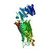 9b8aC  9b8hC  9b8vC M: atomic model generated by this map C: citing same article ( |
|---|---|
| Similar structure data |


-Links
| EMDB pages | EMDB (EBI/PDBe) / EMDataResource |
|---|---|
| Related items in Molecule of the Month |


-Map
| File | Download / File: emd_44346.map.gz / Format: CCP4 / Size: 244.1 MB / Type: IMAGE STORED AS FLOATING POINT NUMBER (4 BYTES) | ||||||||||||||||||||||||||||||||||||
|---|---|---|---|---|---|---|---|---|---|---|---|---|---|---|---|---|---|---|---|---|---|---|---|---|---|---|---|---|---|---|---|---|---|---|---|---|---|
| Annotation | BcsB_BcsC_sharpened map | ||||||||||||||||||||||||||||||||||||
| Projections & slices | Image control
Images are generated by Spider. | ||||||||||||||||||||||||||||||||||||
| Voxel size | X=Y=Z: 1.08 Å | ||||||||||||||||||||||||||||||||||||
| Density |
| ||||||||||||||||||||||||||||||||||||
| Symmetry | Space group: 1 | ||||||||||||||||||||||||||||||||||||
| Details | EMDB XML:
|
 Z (Sec.)
 Y (Row.)
 X (Col.)


-Supplemental data
-Half map: BcsB BcsC halfmap A
| File | emd_44346_half_map_1.map | ||||||||||||
|---|---|---|---|---|---|---|---|---|---|---|---|---|---|
| Annotation | BcsB_BcsC_halfmap_A | ||||||||||||
| Projections & Slices |
| ||||||||||||
| Density Histograms |


-Half map: BcsB BcsC halfmap B
| File | emd_44346_half_map_2.map | ||||||||||||
|---|---|---|---|---|---|---|---|---|---|---|---|---|---|
| Annotation | BcsB_BcsC_halfmap_B | ||||||||||||
| Projections & Slices |
| ||||||||||||
| Density Histograms |


- Sample components
-Entire : Cellulose synthase BcsB-BcsC fusion protein
| Entire | Name: Cellulose synthase BcsB-BcsC fusion protein |
|---|---|
| Components |
|
-Supramolecule #1: Cellulose synthase BcsB-BcsC fusion protein
| Supramolecule | Name: Cellulose synthase BcsB-BcsC fusion protein / type: complex / ID: 1 / Parent: 0 / Macromolecule list: all Details: Proteins: -Bacterial cellulose synthase subunit B (BcsB) -Bacterial cellulose synthase subunit C (BcsC) The BcsC is at the amino-terminus of the construct, so the order is BcsC-linker-BcsB. |
|---|---|
| Source (natural) | Organism: |
-Macromolecule #1: Cellulose synthase BcsB-BcsC fusion protein
| Macromolecule | Name: Cellulose synthase BcsB-BcsC fusion protein / type: protein_or_peptide / ID: 1 Details: N-terminal domain of BcsC (including its first 4 TPRs) was fused to the N-terminus of BcsB via a Gly-Ser linker. The BcsC is at the amino-terminus of the construct, so the order is BcsC-linker-BcsB. Number of copies: 1 / Enantiomer: LEVO |
|---|---|
| Source (natural) | Organism: |
| Molecular weight | Theoretical: 99.242461 KDa |
| Recombinant expression | Organism: |
| Sequence | String: AWSHPQFEKV EAAPTAQQQL LEQVRLGEAT HREDLVQQSL YRLELIDPNN PDVVAARFRS LLRQGDIDGA QKQLDRLSQL APSSNAYKS SRTTMLLSTP DGRQALQQAR LQATTGHAEE AVASYNKLFN GAPPEGDIAV EYWSTVAKIP ARRGEAINQL K RINADAPG ...String: AWSHPQFEKV EAAPTAQQQL LEQVRLGEAT HREDLVQQSL YRLELIDPNN PDVVAARFRS LLRQGDIDGA QKQLDRLSQL APSSNAYKS SRTTMLLSTP DGRQALQQAR LQATTGHAEE AVASYNKLFN GAPPEGDIAV EYWSTVAKIP ARRGEAINQL K RINADAPG GSGSGSGVQG ADAPVVAQNG PSRDVKLTFA QIAPPPGSMV LRGINPNGSI EFGMRSDEVV TKAMLNLEYT PS PSLLPVQ SQLKVYLNDE LMGVLPVTKE QLGKKTLAQM PINPLFISDF NRVRLEFVGH YQDVCEKPAS TTLWLDVGRS SGL DLTYQT LNVKNDLSHF PVPFFDPSDN RTNTLPMVFA GAPDVGLQQA SAIVASWFGS RSGWRGQNFP VLYNQLPDRN AIVF ATNDK RPDFLRDHPA VKAPVIEMIN HPQNPYVKLL VVFGRDDKDL LQAAKGIAQG NILFRGESVV VNEVKPLLPR KPYDA PNWV RTDRPVTFGE LKTYEEQLQS SGLEPAAINV SLNLPPDLYL MRSTGIDMDI NYRYTMPPVK DSSRMDISLN NQFLQS FNL SSKQEANRLL LRIPVLQGLL DGKTDVSIPA LKLGATNQLR FDFEYMNPMP GGSVDNCITF QPVQNHVVIG DDSTIDF SK YYHFIPMPDL RAFANAGFPF SRMADLSQTI TVMPKAPNEA QMETLLNTVG FIGAQTGFPA INLTVTDDGS TIQGKDAD I MIIGGIPDKL KDDKQIDLLV QATESWVKTP MRQTPFPGIV PDESDRAAET RSTLTSSGAM AAVIGFQSPY NDQRSVIAL LADSPRGYEM LNDAVNDSGK RATMFGSVAV IRESGINSLR VGDVYYVGHL PWFERVWYAL ANHPILLAVL AAISVILLAW VLWRLLRII SRRRLNPDNE UniProtKB: Cellulose synthase operon protein C, Cyclic di-GMP-binding protein |
-Experimental details
-Structure determination
| Method | cryo EM |
|---|---|
 Processing Processing | single particle reconstruction |
| Aggregation state | particle |
-Sample preparation
| Buffer | pH: 8 |
|---|---|
| Vitrification | Cryogen name: ETHANE |
- Electron microscopy
| Microscope | FEI TITAN KRIOS |
|---|---|
| Image recording | Film or detector model: GATAN K3 BIOQUANTUM (6k x 4k) / Average electron dose: 50.0 e/Å2 |
| Electron beam | Acceleration voltage: 300 kV / Electron source:  FIELD EMISSION GUN FIELD EMISSION GUN |
| Electron optics | Illumination mode: FLOOD BEAM / Imaging mode: BRIGHT FIELD / Nominal defocus max: 2.0 µm / Nominal defocus min: 1.0 µm |
| Experimental equipment |  Model: Titan Krios / Image courtesy: FEI Company |

