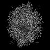
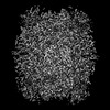
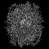
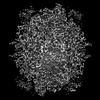
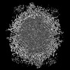
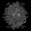
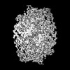
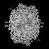
 Movie
Movie Controller

[English] 日本語
 Yorodumi
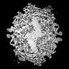
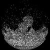
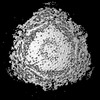
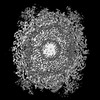
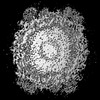
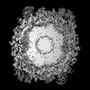
- PDB-9b7n: Fab2-4 in complex with the capsid of Adeno-associated virus type 9 -
+ Open data

- Basic information
| Entry | Database: PDB / ID: 9b7n | |||||||||||||||||||||
|---|---|---|---|---|---|---|---|---|---|---|---|---|---|---|---|---|---|---|---|---|---|---|
| Title | Fab2-4 in complex with the capsid of Adeno-associated virus type 9 | |||||||||||||||||||||
 Components Components |
| |||||||||||||||||||||
 Keywords Keywords | VIRAL PROTEIN/IMMUNE SYSTEM / Adeno-associated virus / AAV / capsid / Fab-complex / antibody / NAb / VIRUS / VIRAL PROTEIN-IMMUNE SYSTEM complex | |||||||||||||||||||||
| Function / homology | Phospholipase A2-like domain / Phospholipase A2-like domain / Parvovirus coat protein VP2 / Parvovirus coat protein VP1/VP2 / Parvovirus coat protein VP1/VP2 / Capsid/spike protein, ssDNA virus / T=1 icosahedral viral capsid / structural molecule activity / Capsid protein VP1 Function and homology information Function and homology information | |||||||||||||||||||||
| Biological species |   Adeno-associated virus Adeno-associated virus Homo sapiens (human) Homo sapiens (human) | |||||||||||||||||||||
| Method | ELECTRON MICROSCOPY / single particle reconstruction / cryo EM / Resolution: 3.02 Å | |||||||||||||||||||||
 Authors Authors | Mietzsch, M. / McKenna, R. | |||||||||||||||||||||
| Funding support |  United States, 1items United States, 1items
| |||||||||||||||||||||
 Citation Citation | Journal: Nat Commun / Year: 2025 Title: Structural characterization of antibody-responses following Zolgensma treatment for AAV capsid engineering to expand patient cohorts. Authors: Mario Mietzsch / Jane Hsi / Austin R Nelson / Neeta Khandekar / Ann-Maree Huang / Nicholas Jc Smith / Jon Zachary / Lindsay Potts / Michelle A Farrar / Paul Chipman / Mohammad Ghanem / Ian E ...Authors: Mario Mietzsch / Jane Hsi / Austin R Nelson / Neeta Khandekar / Ann-Maree Huang / Nicholas Jc Smith / Jon Zachary / Lindsay Potts / Michelle A Farrar / Paul Chipman / Mohammad Ghanem / Ian E Alexander / Grant J Logan / Juha T Huiskonen / Robert McKenna /   Abstract: Monoclonal antibodies are useful tools to dissect the neutralizing antibody response against the adeno-associated virus (AAV) capsids that are used as gene therapy delivery vectors. The presence of ...Monoclonal antibodies are useful tools to dissect the neutralizing antibody response against the adeno-associated virus (AAV) capsids that are used as gene therapy delivery vectors. The presence of pre-existing neutralizing antibodies in large portions of the human population poses a significant challenge for AAV-mediated gene therapy, primarily targeting the capsid leading to vector inactivation and loss of treatment efficacy. This study structurally characterizes the interactions of 21 human-derived neutralizing antibodies from three patients treated with the AAV9 vector, Zolgensma®, utilizing high-resolution cryo-electron microscopy. The antibodies bound to the 2-fold depression or the 3-fold protrusions do not conform to the icosahedral symmetry of the capsid, thus requiring localized reconstructions. These complex structures provide unprecedented details of the mAbs binding interfaces, with many antibodies inducing structural perturbations of the capsid upon binding. Key surface capsid amino acid residues were identified facilitating the design of capsid variants with antibody escape phenotypes. These AAV9 capsid variants have the potential to expand the patient cohort to include those that were previously excluded due to their pre-existing neutralizing antibodies against the wtAAV9 capsid, and the possibly of further treatment to those requiring redosing. | |||||||||||||||||||||
| History |
|
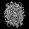
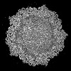
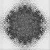
- Structure visualization
| Structure viewer | Molecule: MolmilJmol/JSmol |
|---|
- Downloads & links
-Download
| PDBx/mmCIF format | 9b7n.cif.gz | 459.1 KB | Display | PDBx/mmCIF format |
|---|---|---|---|---|
| PDB format | pdb9b7n.ent.gz | Display | PDB format | |
| PDBx/mmJSON format | 9b7n.json.gz | Tree view | PDBx/mmJSON format | |
| Others |  Other downloads Other downloads |
-Validation report
| Arichive directory | https://data.pdbj.org/pub/pdb/validation_reports/b7/9b7nftp://data.pdbj.org/pub/pdb/validation_reports/b7/9b7n | HTTPS FTP |
|---|
-Related structure data
| Related structure data | 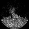 44317MC 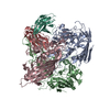 9b6nC 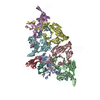 9b6oC 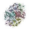 9b6pC 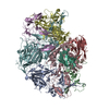 9b6qC 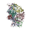 9b6rC 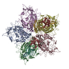 9b6sC  9b6tC 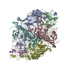 9b7kC 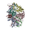 9b7lC 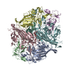 9b7mC 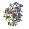 9b7oC 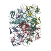 9b7pC 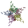 9b7qC 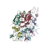 9b7rC 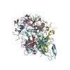 9b7sC 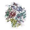 9b7tC 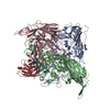 9b7uC 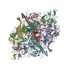 9b7vC  9b7wC 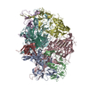 9b7xC M: map data used to model this data C: citing same article ( |
|---|---|
| Similar structure data |
-Links
 PDBj
PDBj


- Assembly
| Deposited unit | 
|
|---|---|
| 1 |
|
-Components
| #1: Protein | Mass: 59873.875 Da / Num. of mol.: 6 Source method: isolated from a genetically manipulated source Source: (gene. exp.) Adeno-associated virus / Strain: AAV9 / Gene: cap / Cell line (production host): HEK293 / Production host: Homo sapiens (human) / References: UniProt: Q6JC22#2: Antibody | | Mass: 11088.250 Da / Num. of mol.: 1 Source method: isolated from a genetically manipulated source Source: (gene. exp.) Homo sapiens (human) / Production host: Homo sapiens (human)#3: Antibody | | Mass: 13834.502 Da / Num. of mol.: 1 Source method: isolated from a genetically manipulated source Source: (gene. exp.) Homo sapiens (human) / Cell line (production host): HEK293 / Production host: Homo sapiens (human)Has protein modification | Y | |
|---|
-Experimental details
-Experiment
| Experiment | Method: ELECTRON MICROSCOPY |
|---|---|
| EM experiment | Aggregation state: PARTICLE / 3D reconstruction method: single particle reconstruction |
- Sample preparation
| Component | Name: Adeno-associated virus / Type: VIRUS / Entity ID: #1 / Source: MULTIPLE SOURCES |
|---|---|
| Source (natural) | Organism: Adeno-associated virus |
| Source (recombinant) | Organism: Homo sapiens (human) / Cell: HEK293 |
| Details of virus | Empty: NO / Enveloped: NO / Isolate: SEROTYPE / Type: VIRION |
| Virus shell | Triangulation number (T number): 1 |
| Buffer solution | pH: 7.4 |
| Specimen | Embedding applied: NO / Shadowing applied: NO / Staining applied: NO / Vitrification applied: YES |
| Vitrification | Cryogen name: ETHANE |
- Electron microscopy imaging
| Experimental equipment |  Model: Titan Krios / Image courtesy: FEI Company |
|---|---|
| Microscopy | Model: TFS KRIOS |
| Electron gun | Electron source:  FIELD EMISSION GUN / Accelerating voltage: 300 kV / Illumination mode: FLOOD BEAM FIELD EMISSION GUN / Accelerating voltage: 300 kV / Illumination mode: FLOOD BEAM |
| Electron lens | Mode: BRIGHT FIELD / Nominal defocus max: 2100 nm / Nominal defocus min: 800 nm |
| Image recording | Electron dose: 50 e/Å2 / Film or detector model: FEI FALCON IV (4k x 4k) |
- Processing
| EM software | Name: PHENIX / Category: model refinement | ||||||||||||||||||||||||
|---|---|---|---|---|---|---|---|---|---|---|---|---|---|---|---|---|---|---|---|---|---|---|---|---|---|
| CTF correction | Type: PHASE FLIPPING AND AMPLITUDE CORRECTION | ||||||||||||||||||||||||
| 3D reconstruction | Resolution: 3.02 Å / Resolution method: FSC 0.143 CUT-OFF / Num. of particles: 360595 / Symmetry type: POINT | ||||||||||||||||||||||||
| Refinement | Stereochemistry target values: REAL-SPACE (WEIGHTED MAP SUM AT ATOM CENTERS) | ||||||||||||||||||||||||
| Refine LS restraints |
|
