Movie
Movie Controller

[English] 日本語
 Yorodumi
- PDB-8z82: Photosynthetic LH1-RC-HiPIP complex from the purple bacterium Hal... -
+ Open data

- Basic information
| Entry | Database: PDB / ID: 8z82 | |||||||||||||||||||||||||||||||||||||||||||||
|---|---|---|---|---|---|---|---|---|---|---|---|---|---|---|---|---|---|---|---|---|---|---|---|---|---|---|---|---|---|---|---|---|---|---|---|---|---|---|---|---|---|---|---|---|---|---|
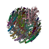
| Title | Photosynthetic LH1-RC-HiPIP complex from the purple bacterium Halorhodospira halophila | |||||||||||||||||||||||||||||||||||||||||||||
 Components Components |
| |||||||||||||||||||||||||||||||||||||||||||||
 Keywords Keywords | PHOTOSYNTHESIS / LH1-RC | |||||||||||||||||||||||||||||||||||||||||||||
| Function / homology |  Function and homology information Function and homology informationorganelle inner membrane / aerobic electron transport chain / plasma membrane-derived chromatophore membrane / plasma membrane light-harvesting complex / bacteriochlorophyll binding / photosynthetic electron transport in photosystem II / photosynthesis, light reaction / endomembrane system / 4 iron, 4 sulfur cluster binding / electron transfer activity ...organelle inner membrane / aerobic electron transport chain / plasma membrane-derived chromatophore membrane / plasma membrane light-harvesting complex / bacteriochlorophyll binding / photosynthetic electron transport in photosystem II / photosynthesis, light reaction / endomembrane system / 4 iron, 4 sulfur cluster binding / electron transfer activity / iron ion binding / heme binding / metal ion binding / plasma membrane Similarity search - Function | |||||||||||||||||||||||||||||||||||||||||||||
| Biological species |  Halorhodospira halophila (bacteria) Halorhodospira halophila (bacteria) | |||||||||||||||||||||||||||||||||||||||||||||
| Method | ELECTRON MICROSCOPY / single particle reconstruction / cryo EM / Resolution: 2.4 Å | |||||||||||||||||||||||||||||||||||||||||||||
 Authors Authors | Tani, K. / Kanno, R. / Nagashima, K.V.P. / Hiwatashi, N. / Kawakami, M. / Nakata, K. / Nagashima, S. / Inoue, K. / Takaichi, S. / Purba, E.R. ...Tani, K. / Kanno, R. / Nagashima, K.V.P. / Hiwatashi, N. / Kawakami, M. / Nakata, K. / Nagashima, S. / Inoue, K. / Takaichi, S. / Purba, E.R. / Hall, M. / Yu, L.-J. / Madigan, M.T. / Mizoguchi, A. / Humbel, B.M. / Kimura, Y. / Wang-Otomo, Z.-Y. | |||||||||||||||||||||||||||||||||||||||||||||
| Funding support |  Japan, 6items Japan, 6items
| |||||||||||||||||||||||||||||||||||||||||||||
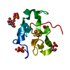
 Citation Citation | Journal: Commun Biol / Year: 2025 Title: A Native LH1-RC-HiPIP Supercomplex from an Extremophilic Phototroph. Authors: Kazutoshi Tani / Ryo Kanno / Kenji V P Nagashima / Mai Kawakami / Naho Hiwatashi / Kazuna Nakata / Sakiko Nagashima / Kazuhito Inoue / Shinichi Takaichi / Endang R Purba / Malgorzata Hall / ...Authors: Kazutoshi Tani / Ryo Kanno / Kenji V P Nagashima / Mai Kawakami / Naho Hiwatashi / Kazuna Nakata / Sakiko Nagashima / Kazuhito Inoue / Shinichi Takaichi / Endang R Purba / Malgorzata Hall / Long-Jiang Yu / Michael T Madigan / Akira Mizoguchi / Bruno M Humbel / Yukihiro Kimura / Zheng-Yu Wang-Otomo /   Abstract: Halorhodospira (Hlr.) halophila strain BN9622 is an extremely halophilic and alkaliphilic purple phototrophic bacterium and has been widely used as a model for exploring the osmoadaptive and ...Halorhodospira (Hlr.) halophila strain BN9622 is an extremely halophilic and alkaliphilic purple phototrophic bacterium and has been widely used as a model for exploring the osmoadaptive and photosynthetic strategies employed by phototrophic extreme halophiles that enable them to thrive in hypersaline environments. Here we present the cryo-EM structures of (1) a unique native Hlr. halophila triple-complex formed from light-harvesting (LH1), the reaction center (RC), and high-potential iron-sulfur protein (HiPIP) at 2.44 Å resolution, and (2) a HiPIP-free LH1-RC complex at 2.64 Å resolution. Differing from the LH1 in the Hlr. halophila LH1-LH2 co-complex where LH1 encircles LH2, the RC-associated LH1 complex consists of 16 (rather than 18) αβ-subunits circularly surrounding the RC. These distinct forms of LH1 indicate that the number of subunits in a Hlr. halophila LH1 complex is flexible and its size is a function of the photocomplex it encircles. Like LH1 in the LH1-LH2 co-complex, the RC-associated LH1 complex also contained two forms of αβ-polypeptides and both dimeric and monomeric molecules of bacteriochlorophyll a. The majority of the isolated Hlr. halophila LH1-RC complexes contained the electron donor HiPIP bound to the surface of the RC cytochrome subunit near the heme-1 group. The bound HiPIP consisted of an N-terminal functional domain and a long C-terminal extension firmly attached to the cytochrome subunit. Despite overall highly negative surface-charge distributions for both the cytochrome subunit and HiPIP, the interface between the two proteins was relatively uncharged and neutral, forming a pathway for electron tunneling. The structure of the Hlr. halophila LH1-RC-HiPIP complex provides insights into the mechanism of light energy acquisition coupled with a long-distance electron donating process toward the charge separation site in a multi-extremophilic phototroph. | |||||||||||||||||||||||||||||||||||||||||||||
| History |
|
- Structure visualization
| Structure viewer | Molecule: MolmilJmol/JSmol |
|---|
- Downloads & links
-Download
| PDBx/mmCIF format | 8z82.cif.gz | 742.4 KB | Display | PDBx/mmCIF format |
|---|---|---|---|---|
| PDB format | pdb8z82.ent.gz | 627.1 KB | Display | PDB format |
| PDBx/mmJSON format | 8z82.json.gz | Tree view | PDBx/mmJSON format | |
| Others |  Other downloads Other downloads |
-Validation report
| Arichive directory | https://data.pdbj.org/pub/pdb/validation_reports/z8/8z82ftp://data.pdbj.org/pub/pdb/validation_reports/z8/8z82 | HTTPS FTP |
|---|
-Related structure data
| Related structure data |  39836MC 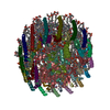 8z83C M: map data used to model this data C: citing same article ( |
|---|---|
| Similar structure data |
-Links
 PDBj
PDBj


- Assembly
| Deposited unit | 
|
|---|---|
| 1 |
|
-Components
-Photosynthetic reaction ... , 2 types, 2 molecules CH
| #1: Protein | Mass: 40439.383 Da / Num. of mol.: 1 / Source method: isolated from a natural source / Source: (natural) Halorhodospira halophila (bacteria) / Strain: BN9622 / References: UniProt: A1WXF5 |
|---|---|
| #4: Protein | Mass: 30904.004 Da / Num. of mol.: 1 / Source method: isolated from a natural source / Source: (natural) Halorhodospira halophila (bacteria) / Strain: BN9622 / References: UniProt: A1WXI3 |
-Reaction center protein ... , 2 types, 2 molecules LM
| #2: Protein | Mass: 30718.809 Da / Num. of mol.: 1 / Source method: isolated from a natural source / Source: (natural) Halorhodospira halophila (bacteria) / Strain: BN9622 / References: UniProt: A0A2L1K3P0 |
|---|---|
| #3: Protein | Mass: 35925.391 Da / Num. of mol.: 1 / Source method: isolated from a natural source / Source: (natural) Halorhodospira halophila (bacteria) / Strain: BN9622 / References: UniProt: A0A2L1K3T5 |
-Antenna complex, alpha/beta ... , 4 types, 32 molecules AFKQUY37BGNRVZ48DIOSW159EJPTX260
| #5: Protein | Mass: 7275.500 Da / Num. of mol.: 8 / Source method: isolated from a natural source / Source: (natural) Halorhodospira halophila (bacteria) / Strain: BN9622 / References: UniProt: A1WWW5#6: Protein | Mass: 8068.154 Da / Num. of mol.: 8 / Source method: isolated from a natural source / Source: (natural) Halorhodospira halophila (bacteria) / Strain: BN9622 / References: UniProt: A1WWW6#7: Protein | Mass: 7664.882 Da / Num. of mol.: 8 / Source method: isolated from a natural source / Source: (natural) Halorhodospira halophila (bacteria) / Strain: BN9622 / References: UniProt: A1WXF8#8: Protein | Mass: 7893.913 Da / Num. of mol.: 8 / Source method: isolated from a natural source / Source: (natural) Halorhodospira halophila (bacteria) / Strain: BN9622 / References: UniProt: A1WXF9 |
|---|
-Protein / Sugars , 2 types, 14 molecules a
| #15: Sugar | ChemComp-LMT /  Type: D-saccharide / Mass: 510.615 Da / Num. of mol.: 13 / Source method: obtained synthetically / Formula: C24H46O11 / Comment: detergent*YM Type: D-saccharide / Mass: 510.615 Da / Num. of mol.: 13 / Source method: obtained synthetically / Formula: C24H46O11 / Comment: detergent*YM#9: Protein | | Mass: 15763.442 Da / Num. of mol.: 1 / Source method: isolated from a natural source / Source: (natural) Halorhodospira halophila (bacteria) / Strain: BN9622 / References: UniProt: A1WXH6 |
|---|
-Non-polymers , 14 types, 125 molecules 


| #10: Chemical | ChemComp-HEC /  Mass: 618.503 Da / Num. of mol.: 4 / Source method: obtained synthetically / Formula: C34H34FeN4O4 Mass: 618.503 Da / Num. of mol.: 4 / Source method: obtained synthetically / Formula: C34H34FeN4O4#11: Chemical | ChemComp-MG / |  Mass: 24.305 Da / Num. of mol.: 1 / Source method: obtained synthetically / Formula: Mg Mass: 24.305 Da / Num. of mol.: 1 / Source method: obtained synthetically / Formula: Mg#12: Chemical | ChemComp-LJQ / [( | Mass: 498.779 Da / Num. of mol.: 1 / Source method: obtained synthetically / Formula: C30H58O5 #13: Chemical | ChemComp-PLM / |  Mass: 256.424 Da / Num. of mol.: 1 / Source method: obtained synthetically / Formula: C16H32O2 Mass: 256.424 Da / Num. of mol.: 1 / Source method: obtained synthetically / Formula: C16H32O2#14: Chemical | ChemComp-PGV / (  Mass: 749.007 Da / Num. of mol.: 42 / Source method: obtained synthetically / Formula: C40H77O10P / Comment: phospholipid*YM Mass: 749.007 Da / Num. of mol.: 42 / Source method: obtained synthetically / Formula: C40H77O10P / Comment: phospholipid*YM#16: Chemical | ChemComp-BCL /  Mass: 911.504 Da / Num. of mol.: 45 / Source method: obtained synthetically / Formula: C55H74MgN4O6 / Feature type: SUBJECT OF INVESTIGATION Mass: 911.504 Da / Num. of mol.: 45 / Source method: obtained synthetically / Formula: C55H74MgN4O6 / Feature type: SUBJECT OF INVESTIGATION#17: Chemical |  Mass: 889.215 Da / Num. of mol.: 2 / Source method: obtained synthetically / Formula: C55H76N4O6 Mass: 889.215 Da / Num. of mol.: 2 / Source method: obtained synthetically / Formula: C55H76N4O6#18: Chemical |  Mass: 727.109 Da / Num. of mol.: 3 / Source method: obtained synthetically / Formula: C49H74O4 Mass: 727.109 Da / Num. of mol.: 3 / Source method: obtained synthetically / Formula: C49H74O4#19: Chemical | ChemComp-FE / |  Mass: 55.845 Da / Num. of mol.: 1 / Source method: obtained synthetically / Formula: Fe Mass: 55.845 Da / Num. of mol.: 1 / Source method: obtained synthetically / Formula: Fe#20: Chemical | ChemComp-CDL /  Mass: 1464.043 Da / Num. of mol.: 9 / Source method: obtained synthetically / Formula: C81H156O17P2 / Comment: phospholipid*YM Mass: 1464.043 Da / Num. of mol.: 9 / Source method: obtained synthetically / Formula: C81H156O17P2 / Comment: phospholipid*YM#21: Chemical | ChemComp-MQ8 / |  Mass: 717.116 Da / Num. of mol.: 1 / Source method: obtained synthetically / Formula: C51H72O2 Mass: 717.116 Da / Num. of mol.: 1 / Source method: obtained synthetically / Formula: C51H72O2#22: Chemical | ChemComp-CRT /  Mass: 596.925 Da / Num. of mol.: 9 / Source method: obtained synthetically / Formula: C42H60O2 Mass: 596.925 Da / Num. of mol.: 9 / Source method: obtained synthetically / Formula: C42H60O2#23: Chemical | ChemComp-SF4 / |  Mass: 351.640 Da / Num. of mol.: 1 / Source method: obtained synthetically / Formula: Fe4S4 Mass: 351.640 Da / Num. of mol.: 1 / Source method: obtained synthetically / Formula: Fe4S4#24: Water | ChemComp-HOH / | Mass: 18.015 Da / Num. of mol.: 5 / Source method: isolated from a natural source / Formula: H2O |
|---|
-Details
| Has ligand of interest | Y |
|---|---|
| Has protein modification | Y |
-Experimental details
-Experiment
| Experiment | Method: ELECTRON MICROSCOPY |
|---|---|
| EM experiment | Aggregation state: PARTICLE / 3D reconstruction method: single particle reconstruction |
- Sample preparation
| Component |
| ||||||||||||||||||
|---|---|---|---|---|---|---|---|---|---|---|---|---|---|---|---|---|---|---|---|
| Molecular weight |
| ||||||||||||||||||
| Source (natural) |
| ||||||||||||||||||
| Buffer solution | pH: 8 | ||||||||||||||||||
| Specimen | Conc.: 6 mg/ml / Embedding applied: NO / Shadowing applied: NO / Staining applied: NO / Vitrification applied: YES / Details: This sample was monodisperse. | ||||||||||||||||||
| Vitrification | Instrument: LEICA EM GP / Cryogen name: ETHANE / Humidity: 90 % / Chamber temperature: 277 K |
- Electron microscopy imaging
| Experimental equipment |  Model: Titan Krios / Image courtesy: FEI Company |
|---|---|
| Microscopy | Model: TFS KRIOS |
| Electron gun | Electron source:  FIELD EMISSION GUN / Accelerating voltage: 300 kV / Illumination mode: FLOOD BEAM FIELD EMISSION GUN / Accelerating voltage: 300 kV / Illumination mode: FLOOD BEAM |
| Electron lens | Mode: BRIGHT FIELD / Nominal defocus max: 2700 nm / Nominal defocus min: 1100 nm / Cs: 2.7 mm / Alignment procedure: COMA FREE |
| Specimen holder | Cryogen: NITROGEN / Specimen holder model: FEI TITAN KRIOS AUTOGRID HOLDER |
| Image recording | Electron dose: 40 e/Å2 / Detector mode: COUNTING / Film or detector model: FEI FALCON III (4k x 4k) |
- Processing
| EM software |
| ||||||||||||||||||||||||||||||||||||||||
|---|---|---|---|---|---|---|---|---|---|---|---|---|---|---|---|---|---|---|---|---|---|---|---|---|---|---|---|---|---|---|---|---|---|---|---|---|---|---|---|---|---|
| CTF correction | Type: PHASE FLIPPING ONLY | ||||||||||||||||||||||||||||||||||||||||
| Particle selection | Num. of particles selected: 331335 | ||||||||||||||||||||||||||||||||||||||||
| 3D reconstruction | Resolution: 2.4 Å / Resolution method: FSC 0.143 CUT-OFF / Num. of particles: 90126 / Algorithm: FOURIER SPACE / Symmetry type: POINT | ||||||||||||||||||||||||||||||||||||||||
| Atomic model building | B value: 20 / Protocol: RIGID BODY FIT / Space: REAL / Target criteria: Correlation coefficient | ||||||||||||||||||||||||||||||||||||||||
| Atomic model building |
| ||||||||||||||||||||||||||||||||||||||||
| Refinement | Highest resolution: 2.4 Å Stereochemistry target values: REAL-SPACE (WEIGHTED MAP SUM AT ATOM CENTERS) | ||||||||||||||||||||||||||||||||||||||||
| Refine LS restraints |
|