Movie
Movie Controller

[English] 日本語
 Yorodumi
- EMDB-39837: Photosynthetic LH1-RC complex from the purple bacterium Halorhodo... -
+ Open data

- Basic information
| Entry |  | |||||||||||||||||||||
|---|---|---|---|---|---|---|---|---|---|---|---|---|---|---|---|---|---|---|---|---|---|---|
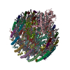
| Title | Photosynthetic LH1-RC complex from the purple bacterium Halorhodospira halophila | |||||||||||||||||||||
 Map data Map data | full map | |||||||||||||||||||||
 Sample Sample |
| |||||||||||||||||||||
 Keywords Keywords | LH1-RC / PHOTOSYNTHESIS | |||||||||||||||||||||
| Function / homology |  Function and homology information Function and homology informationorganelle inner membrane / plasma membrane-derived chromatophore membrane / plasma membrane light-harvesting complex / bacteriochlorophyll binding / photosynthetic electron transport in photosystem II / photosynthesis, light reaction / endomembrane system / metal ion binding / plasma membrane Similarity search - Function | |||||||||||||||||||||
| Biological species |  Halorhodospira halophila (bacteria) Halorhodospira halophila (bacteria) | |||||||||||||||||||||
| Method | single particle reconstruction / cryo EM / Resolution: 2.6 Å | |||||||||||||||||||||
 Authors Authors | Tani K / Kanno R / Nagashima KVP / Hiwatashi N / Kawakami M / Nakata K / Nagashima S / Inoue K / Takaichi S / Purba ER ...Tani K / Kanno R / Nagashima KVP / Hiwatashi N / Kawakami M / Nakata K / Nagashima S / Inoue K / Takaichi S / Purba ER / Hall M / Yu L-J / Madigan MT / Mizoguchi A / Humbel BM / Kimura Y / Wang-Otomo Z-Y | |||||||||||||||||||||
| Funding support |  Japan, 6 items Japan, 6 items
| |||||||||||||||||||||
 Citation Citation | Journal: Commun Biol / Year: 2025 Title: A Native LH1-RC-HiPIP Supercomplex from an Extremophilic Phototroph. Authors: Kazutoshi Tani / Ryo Kanno / Kenji V P Nagashima / Mai Kawakami / Naho Hiwatashi / Kazuna Nakata / Sakiko Nagashima / Kazuhito Inoue / Shinichi Takaichi / Endang R Purba / Malgorzata Hall / ...Authors: Kazutoshi Tani / Ryo Kanno / Kenji V P Nagashima / Mai Kawakami / Naho Hiwatashi / Kazuna Nakata / Sakiko Nagashima / Kazuhito Inoue / Shinichi Takaichi / Endang R Purba / Malgorzata Hall / Long-Jiang Yu / Michael T Madigan / Akira Mizoguchi / Bruno M Humbel / Yukihiro Kimura / Zheng-Yu Wang-Otomo /   Abstract: Halorhodospira (Hlr.) halophila strain BN9622 is an extremely halophilic and alkaliphilic purple phototrophic bacterium and has been widely used as a model for exploring the osmoadaptive and ...Halorhodospira (Hlr.) halophila strain BN9622 is an extremely halophilic and alkaliphilic purple phototrophic bacterium and has been widely used as a model for exploring the osmoadaptive and photosynthetic strategies employed by phototrophic extreme halophiles that enable them to thrive in hypersaline environments. Here we present the cryo-EM structures of (1) a unique native Hlr. halophila triple-complex formed from light-harvesting (LH1), the reaction center (RC), and high-potential iron-sulfur protein (HiPIP) at 2.44 Å resolution, and (2) a HiPIP-free LH1-RC complex at 2.64 Å resolution. Differing from the LH1 in the Hlr. halophila LH1-LH2 co-complex where LH1 encircles LH2, the RC-associated LH1 complex consists of 16 (rather than 18) αβ-subunits circularly surrounding the RC. These distinct forms of LH1 indicate that the number of subunits in a Hlr. halophila LH1 complex is flexible and its size is a function of the photocomplex it encircles. Like LH1 in the LH1-LH2 co-complex, the RC-associated LH1 complex also contained two forms of αβ-polypeptides and both dimeric and monomeric molecules of bacteriochlorophyll a. The majority of the isolated Hlr. halophila LH1-RC complexes contained the electron donor HiPIP bound to the surface of the RC cytochrome subunit near the heme-1 group. The bound HiPIP consisted of an N-terminal functional domain and a long C-terminal extension firmly attached to the cytochrome subunit. Despite overall highly negative surface-charge distributions for both the cytochrome subunit and HiPIP, the interface between the two proteins was relatively uncharged and neutral, forming a pathway for electron tunneling. The structure of the Hlr. halophila LH1-RC-HiPIP complex provides insights into the mechanism of light energy acquisition coupled with a long-distance electron donating process toward the charge separation site in a multi-extremophilic phototroph. | |||||||||||||||||||||
| History |
|
- Structure visualization
| Supplemental images |
|---|

- Downloads & links
-EMDB archive
| Map data | emd_39837.map.gz | 229 MB | EMDB map data format | |
|---|---|---|---|---|
| Header (meta data) | emd-39837-v30.xmlemd-39837.xml | 32.2 KB 32.2 KB | Display Display | EMDB header |
| FSC (resolution estimation) | emd_39837_fsc.xml | 13.6 KB | Display | FSC data file |
| Images |  emd_39837.png emd_39837.png | 130.1 KB | ||
| Filedesc metadata | emd-39837.cif.gz | 8.1 KB | ||
| Others | emd_39837_half_map_1.map.gzemd_39837_half_map_2.map.gz | 194 MB 194.1 MB | ||
| Archive directory |  http://ftp.pdbj.org/pub/emdb/structures/EMD-39837ftp://ftp.pdbj.org/pub/emdb/structures/EMD-39837 http://ftp.pdbj.org/pub/emdb/structures/EMD-39837ftp://ftp.pdbj.org/pub/emdb/structures/EMD-39837 | HTTPS FTP |
-Related structure data
| Related structure data | 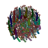 8z83MC 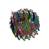 8z82C M: atomic model generated by this map C: citing same article ( |
|---|---|
| Similar structure data |
-Links
| EMDB pages | EMDB (EBI/PDBe) / EMDataResource |
|---|---|
| Related items in Molecule of the Month |


-Map
| File | Download / File: emd_39837.map.gz / Format: CCP4 / Size: 244.1 MB / Type: IMAGE STORED AS FLOATING POINT NUMBER (4 BYTES) | ||||||||||||||||||||||||||||||||||||
|---|---|---|---|---|---|---|---|---|---|---|---|---|---|---|---|---|---|---|---|---|---|---|---|---|---|---|---|---|---|---|---|---|---|---|---|---|---|
| Annotation | full map | ||||||||||||||||||||||||||||||||||||
| Projections & slices | Image control
Images are generated by Spider. | ||||||||||||||||||||||||||||||||||||
| Voxel size | X=Y=Z: 0.82 Å | ||||||||||||||||||||||||||||||||||||
| Density |
| ||||||||||||||||||||||||||||||||||||
| Symmetry | Space group: 1 | ||||||||||||||||||||||||||||||||||||
| Details | EMDB XML:
|
 Z (Sec.)
 Y (Row.)
 X (Col.)


-Supplemental data
-Half map: even-half map
| File | emd_39837_half_map_1.map | ||||||||||||
|---|---|---|---|---|---|---|---|---|---|---|---|---|---|
| Annotation | even-half map | ||||||||||||
| Projections & Slices |
| ||||||||||||
| Density Histograms |


-Half map: odd-half map
| File | emd_39837_half_map_2.map | ||||||||||||
|---|---|---|---|---|---|---|---|---|---|---|---|---|---|
| Annotation | odd-half map | ||||||||||||
| Projections & Slices |
| ||||||||||||
| Density Histograms |


- Sample components
+Entire : Photosynthetic LH1-RC complex from the purple phototrophic bacter...
+Supramolecule #1: Photosynthetic LH1-RC complex from the purple phototrophic bacter...
+Supramolecule #2: LH1-RC complex of Halorhodospira halophila
+Macromolecule #1: Photosynthetic reaction center cytochrome c subunit
+Macromolecule #2: Reaction center protein L chain
+Macromolecule #3: Reaction center protein M chain
+Macromolecule #4: Photosynthetic reaction center H subunit
+Macromolecule #5: Antenna complex, alpha/beta subunit
+Macromolecule #6: Antenna complex, alpha/beta subunit
+Macromolecule #7: Antenna complex, alpha/beta subunit
+Macromolecule #8: Antenna complex, alpha/beta subunit
+Macromolecule #9: HEME C

+Macromolecule #10: MAGNESIUM ION
+Macromolecule #11: (2S)-3-hydroxypropane-1,2-diyl dihexadecanoate
+Macromolecule #12: PALMITIC ACID

+Macromolecule #13: DODECYL-BETA-D-MALTOSIDE

+Macromolecule #14: (1R)-2-{[{[(2S)-2,3-DIHYDROXYPROPYL]OXY}(HYDROXY)PHOSPHORYL]OXY}-...

+Macromolecule #15: BACTERIOCHLOROPHYLL A

+Macromolecule #16: BACTERIOPHEOPHYTIN A

+Macromolecule #17: Ubiquinone-8

+Macromolecule #18: CARDIOLIPIN

+Macromolecule #19: FE (III) ION
+Macromolecule #20: MENAQUINONE 8

+Macromolecule #21: SPIRILLOXANTHIN

+Macromolecule #22: water

-Experimental details
-Structure determination
| Method | cryo EM |
|---|---|
 Processing Processing | single particle reconstruction |
| Aggregation state | particle |
-Sample preparation
| Concentration | 6.0 mg/mL |
|---|---|
| Buffer | pH: 8 |
| Vitrification | Cryogen name: ETHANE / Chamber humidity: 90 % / Chamber temperature: 277 K / Instrument: LEICA EM GP |
| Details | This sample was monodisperse. |
- Electron microscopy
| Microscope | FEI TITAN KRIOS |
|---|---|
| Image recording | Film or detector model: FEI FALCON III (4k x 4k) / Detector mode: COUNTING / Average electron dose: 40.0 e/Å2 |
| Electron beam | Acceleration voltage: 300 kV / Electron source:  FIELD EMISSION GUN FIELD EMISSION GUN |
| Electron optics | Illumination mode: FLOOD BEAM / Imaging mode: BRIGHT FIELD / Cs: 2.7 mm / Nominal defocus max: 2.7 µm / Nominal defocus min: 1.1 µm |
| Sample stage | Specimen holder model: FEI TITAN KRIOS AUTOGRID HOLDER / Cooling holder cryogen: NITROGEN |
| Experimental equipment |  Model: Titan Krios / Image courtesy: FEI Company |
+Image processing

-Atomic model buiding 1
| Initial model | PDB ID: Chain - Source name: PDB / Chain - Initial model type: experimental model |
|---|---|
| Refinement | Space: REAL / Protocol: RIGID BODY FIT / Overall B value: 20 / Target criteria: Correlation coefficient |
| Output model | PDB-8z83: |