 Movie
Movie Controller

[English] 日本語
 Yorodumi
- PDB-8ysl: Crystal structure of the Deinococcus wulumuqiensis CD-NTase DwCdn... -
+ Open data

- Basic information
| Entry | Database: PDB / ID: 8ysl | ||||||
|---|---|---|---|---|---|---|---|
| Title | Crystal structure of the Deinococcus wulumuqiensis CD-NTase DwCdnB in complex with UTP and ATP | ||||||
 Components Components | Nucleotidyltransferase | ||||||
 Keywords Keywords | TRANSFERASE / CD-NTase | ||||||
| Function / homology |  Function and homology information Function and homology informationnucleotide metabolic process / nucleotidyltransferase activity / defense response to virus / GTP binding / ATP binding / metal ion binding Similarity search - Function | ||||||
| Biological species |  Deinococcus wulumuqiensis (bacteria) Deinococcus wulumuqiensis (bacteria) | ||||||
| Method |  X-RAY DIFFRACTION / SYNCHROTRON / MOLECULAR REPLACEMENT / Resolution: 1.98 Å X-RAY DIFFRACTION / SYNCHROTRON / MOLECULAR REPLACEMENT / Resolution: 1.98 Å | ||||||
 Authors Authors | Wang, Y.-C. / Yang, C.-S. / Hou, M.-H. / Chen, Y. | ||||||
| Funding support |  Taiwan, 1items Taiwan, 1items
| ||||||
 Citation Citation | Journal: Int.J.Biol.Macromol. / Year: 2025 Title: Structural insights into signaling promiscuity of the CBASS anti-phage defense system from a radiation-resistant bacterium. Authors: Yang, C.S. / Shie, M.Y. / Huang, S.W. / Wang, Y.C. / Hou, M.H. / Chen, C.J. / Chen, Y. | ||||||
| History |
|
- Structure visualization
| Structure viewer | Molecule: MolmilJmol/JSmol |
|---|
- Downloads & links
-Download
| PDBx/mmCIF format | 8ysl.cif.gz | 156.3 KB | Display | PDBx/mmCIF format |
|---|---|---|---|---|
| PDB format | pdb8ysl.ent.gz | 120.1 KB | Display | PDB format |
| PDBx/mmJSON format | 8ysl.json.gz | Tree view | PDBx/mmJSON format | |
| Others |  Other downloads Other downloads |
-Validation report
| Arichive directory | https://data.pdbj.org/pub/pdb/validation_reports/ys/8yslftp://data.pdbj.org/pub/pdb/validation_reports/ys/8ysl | HTTPS FTP |
|---|
-Related structure data
| Related structure data |  8yscC  8ysdC  8yseC  8ysgC  8ysiC  8ysjC  8yskC  8ysmC  8ysnC  8ysoC 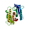 8ysqC  8ysrC  8yssC  8ysuC C: citing same article ( |
|---|---|
| Similar structure data |
-Links
 PDBj
PDBj- Assembly
| Deposited unit | 
| ||||||||
|---|---|---|---|---|---|---|---|---|---|
| 1 |
| ||||||||
| Unit cell |
|
-Components
| #1: Protein | Mass: 39349.109 Da / Num. of mol.: 1 Source method: isolated from a genetically manipulated source Details: The first residue should be annotated as Met0 / Source: (gene. exp.) Deinococcus wulumuqiensis (bacteria) / Gene: DVJ83_15700 / Production host: | ||||||
|---|---|---|---|---|---|---|---|
| #2: Chemical | ChemComp-ATP /   Mass: 507.181 Da / Num. of mol.: 1 / Source method: obtained synthetically / Formula: C10H16N5O13P3 / Feature type: SUBJECT OF INVESTIGATION / Comment: ATP, energy-carrying molecule*YM Mass: 507.181 Da / Num. of mol.: 1 / Source method: obtained synthetically / Formula: C10H16N5O13P3 / Feature type: SUBJECT OF INVESTIGATION / Comment: ATP, energy-carrying molecule*YM | ||||||
| #3: Chemical | ChemComp-UTP /   Mass: 484.141 Da / Num. of mol.: 1 / Source method: obtained synthetically / Formula: C9H15N2O15P3 / Feature type: SUBJECT OF INVESTIGATION / Comment: UTP*YM Mass: 484.141 Da / Num. of mol.: 1 / Source method: obtained synthetically / Formula: C9H15N2O15P3 / Feature type: SUBJECT OF INVESTIGATION / Comment: UTP*YM | ||||||
| #4: Chemical |   Mass: 24.305 Da / Num. of mol.: 2 / Source method: obtained synthetically / Formula: Mg Mass: 24.305 Da / Num. of mol.: 2 / Source method: obtained synthetically / Formula: Mg#5: Water | ChemComp-HOH / |  Mass: 18.015 Da / Num. of mol.: 288 / Source method: isolated from a natural source / Formula: H2O Mass: 18.015 Da / Num. of mol.: 288 / Source method: isolated from a natural source / Formula: H2OHas ligand of interest | Y | Has protein modification | N | |
-Experimental details
-Experiment
| Experiment | Method: X-RAY DIFFRACTION / Number of used crystals: 1 |
|---|
- Sample preparation
| Crystal | Density Matthews: 2.31 Å3/Da / Density % sol: 46.71 % |
|---|---|
| Crystal grow | Temperature: 277 K / Method: vapor diffusion, sitting drop Details: 0.1 M Potassium thiocyanate, 30% w/v Polyethylene glycol monomethyl ether 2,000. |
-Data collection
| Diffraction | Mean temperature: 100 K / Serial crystal experiment: N |
|---|---|
| Diffraction source | Source: SYNCHROTRON / Site: NSRRC / Beamline: TPS 05A / Wavelength: 1 Å |
| Detector | Type: DECTRIS EIGER2 X 9M / Detector: PIXEL / Date: Mar 26, 2021 |
| Radiation | Protocol: SINGLE WAVELENGTH / Monochromatic (M) / Laue (L): M / Scattering type: x-ray |
| Radiation wavelength | Wavelength: 1 Å / Relative weight: 1 |
| Reflection | Resolution: 1.98→30 Å / Num. obs: 23777 / % possible obs: 97 % / Redundancy: 2.4 % / Rmerge(I) obs: 0.042 / Net I/σ(I): 20.293 |
| Reflection shell | Resolution: 1.98→2.05 Å / Num. unique obs: 2356 / CC1/2: 0.946 |
- Processing
| Software |
| ||||||||||||||||
|---|---|---|---|---|---|---|---|---|---|---|---|---|---|---|---|---|---|
| Refinement | Method to determine structure: MOLECULAR REPLACEMENT / Resolution: 1.98→26.59 Å / Cross valid method: NONE
| ||||||||||||||||
| Refinement step | Cycle: LAST / Resolution: 1.98→26.59 Å
|
