 Movie
Movie Controller

[English] 日本語
 Yorodumi
- PDB-8ypz: Crystal strcture of human phosphoribosyl pyrophosphate synthetase... -
+ Open data

- Basic information
| Entry | Database: PDB / ID: 8ypz | ||||||
|---|---|---|---|---|---|---|---|
| Title | Crystal strcture of human phosphoribosyl pyrophosphate synthetase 1 (PRPS1) in complex with GDP | ||||||
 Components Components | Ribose-phosphate pyrophosphokinase 1 | ||||||
 Keywords Keywords | BIOSYNTHETIC PROTEIN / Purine biosynthesis | ||||||
| Function / homology |  Function and homology information Function and homology information5-Phosphoribose 1-diphosphate biosynthesis / hypoxanthine biosynthetic process / ribose phosphate diphosphokinase complex / ribonucleoside monophosphate biosynthetic process / ribose-phosphate diphosphokinase / ribose phosphate diphosphokinase activity / urate biosynthetic process / 5-phosphoribose 1-diphosphate biosynthetic process / pyrimidine nucleotide biosynthetic process / purine nucleotide biosynthetic process ...5-Phosphoribose 1-diphosphate biosynthesis / hypoxanthine biosynthetic process / ribose phosphate diphosphokinase complex / ribonucleoside monophosphate biosynthetic process / ribose-phosphate diphosphokinase / ribose phosphate diphosphokinase activity / urate biosynthetic process / 5-phosphoribose 1-diphosphate biosynthetic process / pyrimidine nucleotide biosynthetic process / purine nucleotide biosynthetic process / purine nucleobase metabolic process / kinase activity / nervous system development / magnesium ion binding / protein homodimerization activity / ATP binding / identical protein binding / cytoplasm / cytosol Similarity search - Function | ||||||
| Biological species |  Homo sapiens (human) Homo sapiens (human) | ||||||
| Method |  X-RAY DIFFRACTION / SYNCHROTRON / MOLECULAR REPLACEMENT / Resolution: 3 Å X-RAY DIFFRACTION / SYNCHROTRON / MOLECULAR REPLACEMENT / Resolution: 3 Å | ||||||
 Authors Authors | Zhang, L. / Zhang, L. | ||||||
| Funding support |  China, 1items China, 1items
| ||||||
 Citation Citation | Journal: Nat Commun / Year: 2025 Title: PRPS2 enhances RNA m 6 A methylation by stimulating SAM synthesis through enzyme-dependent and independent mechanisms. Authors: Zhang, L. / Zhao, X. / Hu, J. / Li, T. / Chen, H.Z. / Zhang, A. / Wang, H. / Yu, J. / Zhang, L. | ||||||
| History |
|
- Structure visualization
| Structure viewer | Molecule: MolmilJmol/JSmol |
|---|
- Downloads & links
-Download
| PDBx/mmCIF format | 8ypz.cif.gz | 363.3 KB | Display | PDBx/mmCIF format |
|---|---|---|---|---|
| PDB format | pdb8ypz.ent.gz | 296.8 KB | Display | PDB format |
| PDBx/mmJSON format | 8ypz.json.gz | Tree view | PDBx/mmJSON format | |
| Others |  Other downloads Other downloads |
-Validation report
| Arichive directory | https://data.pdbj.org/pub/pdb/validation_reports/yp/8ypzftp://data.pdbj.org/pub/pdb/validation_reports/yp/8ypz | HTTPS FTP |
|---|
-Related structure data
| Related structure data |  8ypyC 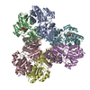 8yq0C 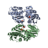 2h06S S: Starting model for refinement C: citing same article ( |
|---|---|
| Similar structure data |
-Links
 PDBj
PDBj


- Assembly
| Deposited unit | 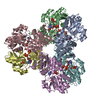
| ||||||||
|---|---|---|---|---|---|---|---|---|---|
| 1 |
| ||||||||
| Unit cell |
|
-Components
-Protein / Sugars , 2 types, 11 molecules ABCDEF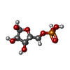
| #1: Protein | Mass: 34835.121 Da / Num. of mol.: 6 Source method: isolated from a genetically manipulated source Source: (gene. exp.) Homo sapiens (human) / Gene: PRPS1 / Production host:  References: UniProt: P60891, ribose-phosphate diphosphokinase #3: Sugar | ChemComp-HSX /  Type: D-saccharide, alpha linking / Mass: 230.110 Da / Num. of mol.: 5 / Source method: obtained synthetically / Formula: C5H11O8P Type: D-saccharide, alpha linking / Mass: 230.110 Da / Num. of mol.: 5 / Source method: obtained synthetically / Formula: C5H11O8P |
|---|
-Non-polymers , 4 types, 81 molecules 


| #2: Chemical | ChemComp-APC /  Mass: 505.208 Da / Num. of mol.: 6 / Source method: obtained synthetically / Formula: C11H18N5O12P3 / Comment: AMP-CPP, energy-carrying molecule analogue*YM Mass: 505.208 Da / Num. of mol.: 6 / Source method: obtained synthetically / Formula: C11H18N5O12P3 / Comment: AMP-CPP, energy-carrying molecule analogue*YM#4: Chemical | ChemComp-GDP /  Type: RNA linking / Mass: 443.201 Da / Num. of mol.: 6 / Source method: obtained synthetically / Formula: C10H15N5O11P2 / Feature type: SUBJECT OF INVESTIGATION / Comment: GDP, energy-carrying molecule*YM Type: RNA linking / Mass: 443.201 Da / Num. of mol.: 6 / Source method: obtained synthetically / Formula: C10H15N5O11P2 / Feature type: SUBJECT OF INVESTIGATION / Comment: GDP, energy-carrying molecule*YM#5: Chemical | ChemComp-PO4 /  Mass: 94.971 Da / Num. of mol.: 6 / Source method: obtained synthetically / Formula: PO4 Mass: 94.971 Da / Num. of mol.: 6 / Source method: obtained synthetically / Formula: PO4#6: Water | ChemComp-HOH / | Mass: 18.015 Da / Num. of mol.: 63 / Source method: isolated from a natural source / Formula: H2O |
|---|
-Details
| Has ligand of interest | Y |
|---|---|
| Has protein modification | N |
-Experimental details
-Experiment
| Experiment | Method: X-RAY DIFFRACTION / Number of used crystals: 1 |
|---|
- Sample preparation
| Crystal | Density Matthews: 3.87 Å3/Da / Density % sol: 68.25 % |
|---|---|
| Crystal grow | Temperature: 277 K / Method: vapor diffusion, sitting drop / Details: 0.2M sodium acetate, 10% PEG 3350 |
-Data collection
| Diffraction | Mean temperature: 100 K / Serial crystal experiment: N |
|---|---|
| Diffraction source | Source: SYNCHROTRON / Site: SSRF / Beamline: BL19U1 / Wavelength: 0.9873 Å |
| Detector | Type: DECTRIS PILATUS 6M / Detector: PIXEL / Date: Nov 18, 2018 |
| Radiation | Protocol: SINGLE WAVELENGTH / Monochromatic (M) / Laue (L): M / Scattering type: x-ray |
| Radiation wavelength | Wavelength: 0.9873 Å / Relative weight: 1 |
| Reflection | Resolution: 3→48.7 Å / Num. obs: 62270 / % possible obs: 100 % / Redundancy: 17.4 % / CC1/2: 0.764 / Net I/σ(I): 11.2 |
| Reflection shell | Resolution: 3→3.11 Å / Num. unique obs: 6340 / CC1/2: 0.764 |
- Processing
| Software |
| ||||||||||||||||||
|---|---|---|---|---|---|---|---|---|---|---|---|---|---|---|---|---|---|---|---|
| Refinement | Method to determine structure: MOLECULAR REPLACEMENT Starting model: 2H06 Resolution: 3→48.7 Å / Cross valid method: FREE R-VALUE
| ||||||||||||||||||
| Displacement parameters | Biso mean: 42.05 Å2 | ||||||||||||||||||
| Refinement step | Cycle: LAST / Resolution: 3→48.7 Å
| ||||||||||||||||||
| LS refinement shell | Resolution: 3→3.11 Å
|
