 Movie
Movie Controller


+ Open data

- Basic information
| Entry | Database: PDB / ID: 8xr4 | ||||||
|---|---|---|---|---|---|---|---|
| Title | Crystal structure of AKRtyl-NADP(H) complex | ||||||
 Components Components | Aldo/keto reductase | ||||||
 Keywords Keywords | OXIDOREDUCTASE | ||||||
| Function / homology | : / NADP-dependent oxidoreductase domain / Aldo/keto reductase family / NADP-dependent oxidoreductase domain superfamily / oxidoreductase activity / nucleotide binding / cytosol / NADP NICOTINAMIDE-ADENINE-DINUCLEOTIDE PHOSPHATE / Aldo/keto reductase Function and homology information Function and homology information | ||||||
| Biological species |  Streptomyces xinghaiensis (bacteria) Streptomyces xinghaiensis (bacteria) | ||||||
| Method |  X-RAY DIFFRACTION / SYNCHROTRON / MOLECULAR REPLACEMENT / Resolution: 1.94 Å X-RAY DIFFRACTION / SYNCHROTRON / MOLECULAR REPLACEMENT / Resolution: 1.94 Å | ||||||
 Authors Authors | Lin, S. / Dai, S. / Xiao, Z. | ||||||
| Funding support |  China, 1items China, 1items
| ||||||
 Citation Citation | Journal: Nat Commun / Year: 2024 Title: A three-level regulatory mechanism of the aldo-keto reductase subfamily AKR12D. Authors: Xiao, Z. / Zha, J. / Yang, X. / Huang, T. / Huang, S. / Liu, Q. / Wang, X. / Zhong, J. / Zheng, J. / Liang, R. / Deng, Z. / Zhang, J. / Lin, S. / Dai, S. | ||||||
| History |
|
- Structure visualization
| Structure viewer | Molecule: MolmilJmol/JSmol |
|---|
- Downloads & links
-Download
| PDBx/mmCIF format | 8xr4.cif.gz | 564.9 KB | Display | PDBx/mmCIF format |
|---|---|---|---|---|
| PDB format | pdb8xr4.ent.gz | 460.5 KB | Display | PDB format |
| PDBx/mmJSON format | 8xr4.json.gz | Tree view | PDBx/mmJSON format | |
| Others |  Other downloads Other downloads |
-Validation report
| Arichive directory | https://data.pdbj.org/pub/pdb/validation_reports/xr/8xr4ftp://data.pdbj.org/pub/pdb/validation_reports/xr/8xr4 | HTTPS FTP |
|---|
-Related structure data
| Related structure data |  8jwkC  8jwlC 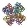 8jwmC  8jwnC  8jwoC  8xr2C 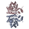 8xr3C C: citing same article ( |
|---|---|
| Similar structure data |
-Links
 PDBj
PDBj
- Assembly
| Deposited unit | 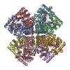
| ||||||||||||
|---|---|---|---|---|---|---|---|---|---|---|---|---|---|
| 1 |
| ||||||||||||
| Unit cell |
|
-Components
| #1: Protein | Mass: 36293.703 Da / Num. of mol.: 8 Source method: isolated from a genetically manipulated source Source: (gene. exp.) Streptomyces xinghaiensis (bacteria) / Gene: DC095_024450, SFRA_012675 / Production host: #2: Chemical | ChemComp-NAP /   Mass: 743.405 Da / Num. of mol.: 8 / Source method: obtained synthetically / Formula: C21H28N7O17P3 / Feature type: SUBJECT OF INVESTIGATION Mass: 743.405 Da / Num. of mol.: 8 / Source method: obtained synthetically / Formula: C21H28N7O17P3 / Feature type: SUBJECT OF INVESTIGATION#3: Water | ChemComp-HOH / |  Mass: 18.015 Da / Num. of mol.: 2355 / Source method: isolated from a natural source / Formula: H2O Mass: 18.015 Da / Num. of mol.: 2355 / Source method: isolated from a natural source / Formula: H2OHas ligand of interest | Y | |
|---|
-Experimental details
-Experiment
| Experiment | Method: X-RAY DIFFRACTION / Number of used crystals: 1 |
|---|
- Sample preparation
| Crystal | Density Matthews: 2.77 Å3/Da / Density % sol: 55.52 % |
|---|---|
| Crystal grow | Temperature: 293.15 K / Method: vapor diffusion, sitting drop / Details: 18% PEG3350, 0.1 M Sodium citrate, pH 5.5 |
-Data collection
| Diffraction | Mean temperature: 100 K / Serial crystal experiment: N |
|---|---|
| Diffraction source | Source: SYNCHROTRON / Site: SSRF / Beamline: BL18U1 / Wavelength: 0.97862 Å |
| Detector | Type: DECTRIS PILATUS3 6M / Detector: PIXEL / Date: Dec 3, 2023 |
| Radiation | Monochromator: Si111 / Protocol: SINGLE WAVELENGTH / Monochromatic (M) / Laue (L): M / Scattering type: x-ray |
| Radiation wavelength | Wavelength: 0.97862 Å / Relative weight: 1 |
| Reflection | Resolution: 1.94→45.98 Å / Num. obs: 242437 / % possible obs: 100 % / Redundancy: 12.1 % / Biso Wilson estimate: 26.03 Å2 / CC1/2: 0.996 / Net I/σ(I): 9.6 |
| Reflection shell | Resolution: 1.94→1.97 Å / Mean I/σ(I) obs: 2.1 / Num. unique obs: 11892 / CC1/2: 0.82 |
- Processing
| Software |
| |||||||||||||||||||||||||||||||||||||||||||||||||||||||||||||||||||||||||||||||||||||||||||||||||||||||||||||||||||||||||||||||||||||||||||||||||||||||||||||||||||||||||||||||||||||||||||||||||||||||||||||||||||||||||
|---|---|---|---|---|---|---|---|---|---|---|---|---|---|---|---|---|---|---|---|---|---|---|---|---|---|---|---|---|---|---|---|---|---|---|---|---|---|---|---|---|---|---|---|---|---|---|---|---|---|---|---|---|---|---|---|---|---|---|---|---|---|---|---|---|---|---|---|---|---|---|---|---|---|---|---|---|---|---|---|---|---|---|---|---|---|---|---|---|---|---|---|---|---|---|---|---|---|---|---|---|---|---|---|---|---|---|---|---|---|---|---|---|---|---|---|---|---|---|---|---|---|---|---|---|---|---|---|---|---|---|---|---|---|---|---|---|---|---|---|---|---|---|---|---|---|---|---|---|---|---|---|---|---|---|---|---|---|---|---|---|---|---|---|---|---|---|---|---|---|---|---|---|---|---|---|---|---|---|---|---|---|---|---|---|---|---|---|---|---|---|---|---|---|---|---|---|---|---|---|---|---|---|---|---|---|---|---|---|---|---|---|---|---|---|---|---|---|---|
| Refinement | Method to determine structure: MOLECULAR REPLACEMENT / Resolution: 1.94→45.98 Å / SU ML: 0.2206 / Cross valid method: FREE R-VALUE / σ(F): 1.35 / Phase error: 20.2625 Stereochemistry target values: GeoStd + Monomer Library + CDL v1.2
| |||||||||||||||||||||||||||||||||||||||||||||||||||||||||||||||||||||||||||||||||||||||||||||||||||||||||||||||||||||||||||||||||||||||||||||||||||||||||||||||||||||||||||||||||||||||||||||||||||||||||||||||||||||||||
| Solvent computation | Shrinkage radii: 0.9 Å / VDW probe radii: 1.11 Å / Solvent model: FLAT BULK SOLVENT MODEL | |||||||||||||||||||||||||||||||||||||||||||||||||||||||||||||||||||||||||||||||||||||||||||||||||||||||||||||||||||||||||||||||||||||||||||||||||||||||||||||||||||||||||||||||||||||||||||||||||||||||||||||||||||||||||
| Displacement parameters | Biso mean: 29.31 Å2 | |||||||||||||||||||||||||||||||||||||||||||||||||||||||||||||||||||||||||||||||||||||||||||||||||||||||||||||||||||||||||||||||||||||||||||||||||||||||||||||||||||||||||||||||||||||||||||||||||||||||||||||||||||||||||
| Refinement step | Cycle: LAST / Resolution: 1.94→45.98 Å
| |||||||||||||||||||||||||||||||||||||||||||||||||||||||||||||||||||||||||||||||||||||||||||||||||||||||||||||||||||||||||||||||||||||||||||||||||||||||||||||||||||||||||||||||||||||||||||||||||||||||||||||||||||||||||
| Refine LS restraints |
| |||||||||||||||||||||||||||||||||||||||||||||||||||||||||||||||||||||||||||||||||||||||||||||||||||||||||||||||||||||||||||||||||||||||||||||||||||||||||||||||||||||||||||||||||||||||||||||||||||||||||||||||||||||||||
| LS refinement shell |
|
